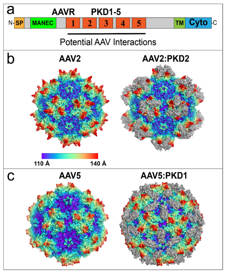
Figure 4.
AAV2 and AAV5 bind to distinct AAVR PKD domains. (a) AAVR domain structure from N-terminus (N) to C-terminus (C): Signal Peptide (SP), Motif At the N-terminus with Eight Cysteines (MANEC), immunoglobulin-like Polycystic Kidney Disease (PKD) domains 1–5, TransMembrane (TM) helix and Cytoplasmic domain (Cyto). The region containing potential AAV interactions is composed of PKD1-5. (b) Native AAV2 60-mer (left) and the AAV2:PKD2 complex (right). Virus models are colored by radial distance from the center of the virion. The PKD2 domain of AAVR is colored in gray. (c) Virus model of native AAV5 virion (left) and the AAV5:PKD1 complex (right). Models are colored by radial distance from the center of the virion. The PKD1 domain of AAVR is colored in gray. Structures in (b,c) were prepared using PyMOL [66].

