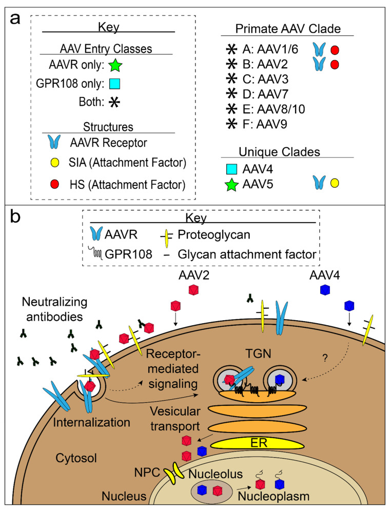
Figure 5.
AAV entry model. (a) Three known AAV entry classes are shown: AAVR-dependent (green star), GPR108-dependent (light blue box) and both AAVR and GPR108-dependent (black asterisk). Structures for AAV complexes with glycan attachment factors (yellow and red circles) and AAV:AAVR complexes (light blue receptor icon) are known. Appropriate AAV entry class icons are located to the left of AAV serotypes and structural icons are located to the right of AAV serotypes with structural evidence. (b) AAV cell attachment, entry, and trafficking to the nucleus of a non-polarized cell. Two AAV classes are shown: AAVR/GPR108-dependent (red; majority of AAVs), represented by AAV2, and GPR108-dependent/AAVR-independent (blue; AAV4 clade). Virions must evade host neutralizing antibodies (black antibody-shaped icons). AAV virions (red or blue) come into contact with the cell surface and attach to the glycan moieties of proteoglycans. Red AAV binds to the AAVR receptor and is internalized and transported through the trans-Golgi network (TGN). Blue, GPR108-dependent, AAV particles enter through a parallel but possibly distinct pathway. Virions accumulate outside of the nucleus before entry and may involve the nuclear pore complex (NPC). AAV gathers in the nucleolus prior to being extruded into the nucleoplasm where the ssDNA genome is released.

