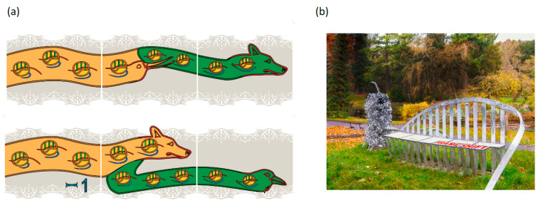
Figure 1.
Outreach representations of frameshifting and readthrough. Panel (a) depicts retroviral recoding where protease sequencing was important for mechanistic understanding: top, Murine Leukemia Virus gag stop codon Readthrough; bottom, Mouse Mammary Tumor Virus gag pol Frameshifting. The head motifs used to depict directionality were inspired by the 1200-year old ‘Book of Kells’ in Trinity College Dublin library. The embedded ribosomes have their A, P, and E sites in green and those shown on the left are at an earlier stage of decoding than those on the right. Correspondingly, on the left the proportion of the mRNA (in red) that has passed through the ribosomes is small in contrast to that shown on the right side, and the nascent peptide emerging from the ribosome (blue) is longer on the right side than on the left. Gag is represented in ochre and Pol in green. These images are from a band of recoding tiles positioned around the middle of the outside walls of ‘a house’ in S.W. Cork, Ireland. Panel (b) shows the decoding seat component of the sculpture in the National Botanic Gardens, Dublin entitled ‘What is Life’. The title follows that used by Erwin Schrödinger of the Dublin Institute of Advanced Studies for his 1944 book (and previous year lectures). Both Watson and Crick independently credited the book ‘What is Life?’ as an early source of inspiration for them. A description of the components of the sculpture, including a hammerhead ribozyme and a ribosome can be found at http://whatislife.ie/ (accessed on 1 May 2021). The decoding seat is on a mound overlooking an iconic Charles Jencks 5.5 m high DNA double helix similar to those at Clare College Cambridge and near Cold Spring Harbor Laboratory beach. Each seat panel represents a codon. Three bars below and above each panel reflect its 3nt composition. Starting from the left, or 5′ end, the initial panels reflect all zero frame reading. A proportion of ribosomes shifting to the -1 frame is represented by the first split panel in which part of the panel is offset to the left by one third of a panel length. Continued triplet decoding by frameshifted ribosomes, and by zero frame ribosomes is represented by the panel at the right end.

