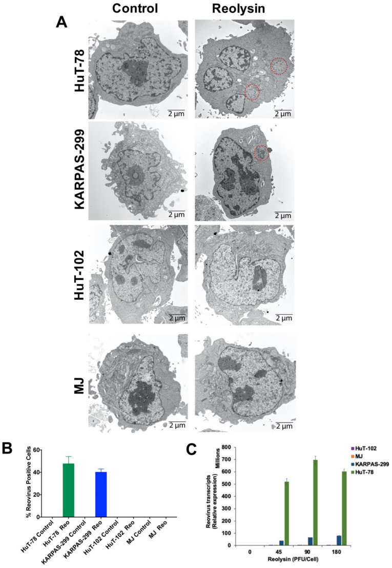
Figure 2.
HuT-78 and KARPAS-299 TCL cells are sensitive to oncolytic reovirus replication. (A) Oncolytic reovirus replicates in HuT-78 and KARPAS-299 cells. HuT-78, KARPAS-299, HuT-102 and MJ cells were treated with 90 PFU/cell Reolysin for 48 h. Reovirus accumulation was visualized by transmission electron microscopy. Red circles indicate reovirus accumulation. Representative images are shown. (B) Quantification of reovirus in TCL cells. The percentage of reovirus-positive cells was counted in approximately 200 cells per cell line using transmission electron microscopy. No reovirus-positive cells were observed in the HTLV-1-positive HuT-102 and MJ cell lines. Mean ± SD. (C) Quantification of reovirus transcripts by qRT-PCR. TCL cell lines were treated with the indicated concentrations of Reolysin for 48 h. Relative levels of reovirus were determined by comparing 48 h Reolysin-treated samples with Controls for each cell line by qRT-PCR. Mean ± SD, n = 3.

