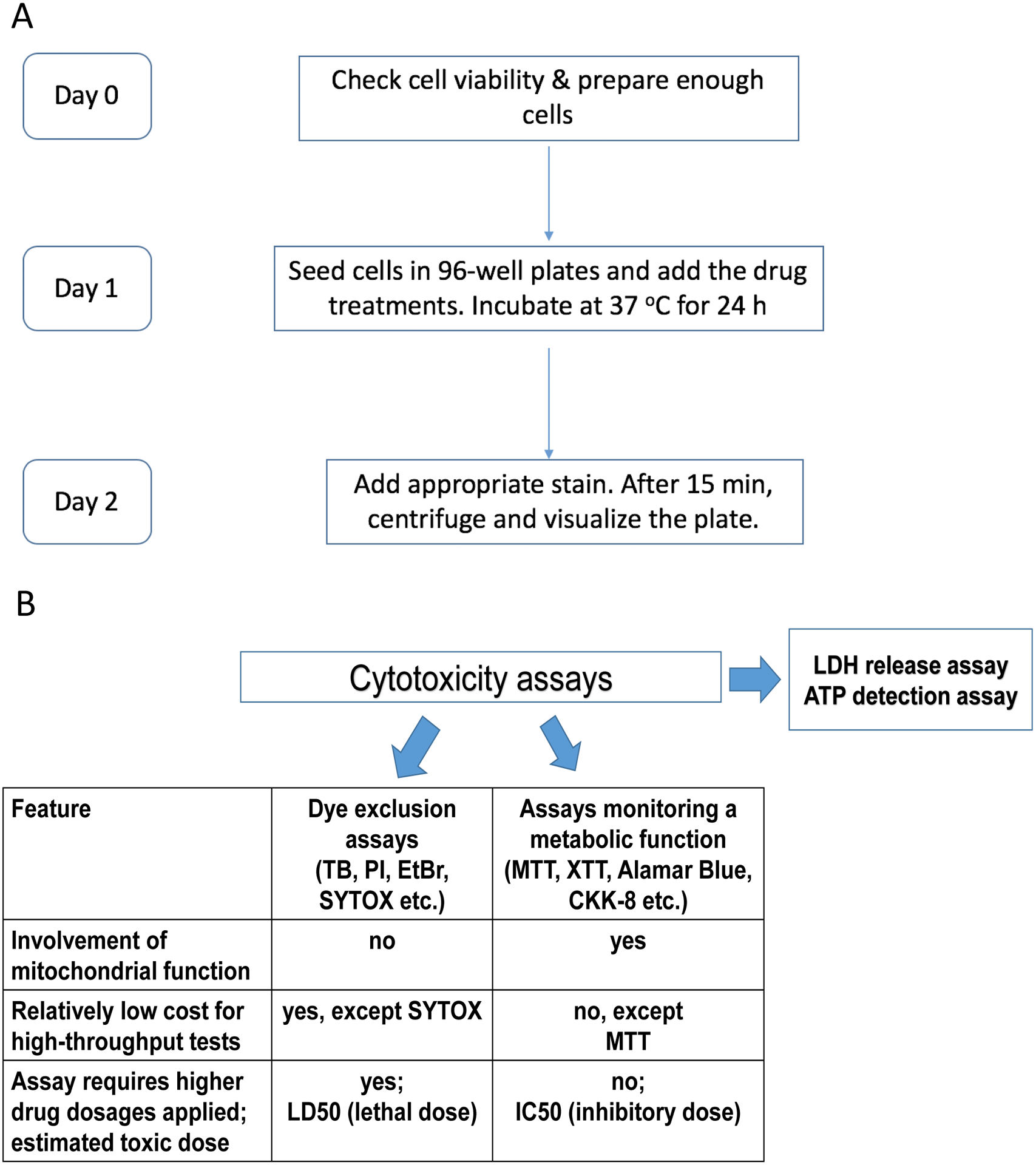
Figure 1. Experimental timeline and comparison of existing cytotoxicity assays.

A. Flowchart summarizing the timeline for the experimental procedure, e.g. Hoechst/PI staining. B. Comparison of cytotoxicity assays, some of which were used in this study. Dye exclusion assays involve impermeant nuclear dyes that stain dead cells with compromised plasma membrane: TB – trypan blue, PI – propidium iodide, EtBr – ethidium bromide, and SYTOX. Second group of assays depend on cellular metabolism, for example, tetrazolium salts MTT, XTT, and CKK-8 (WST-8), resazurin-based reagent alamarBlue, etc.
