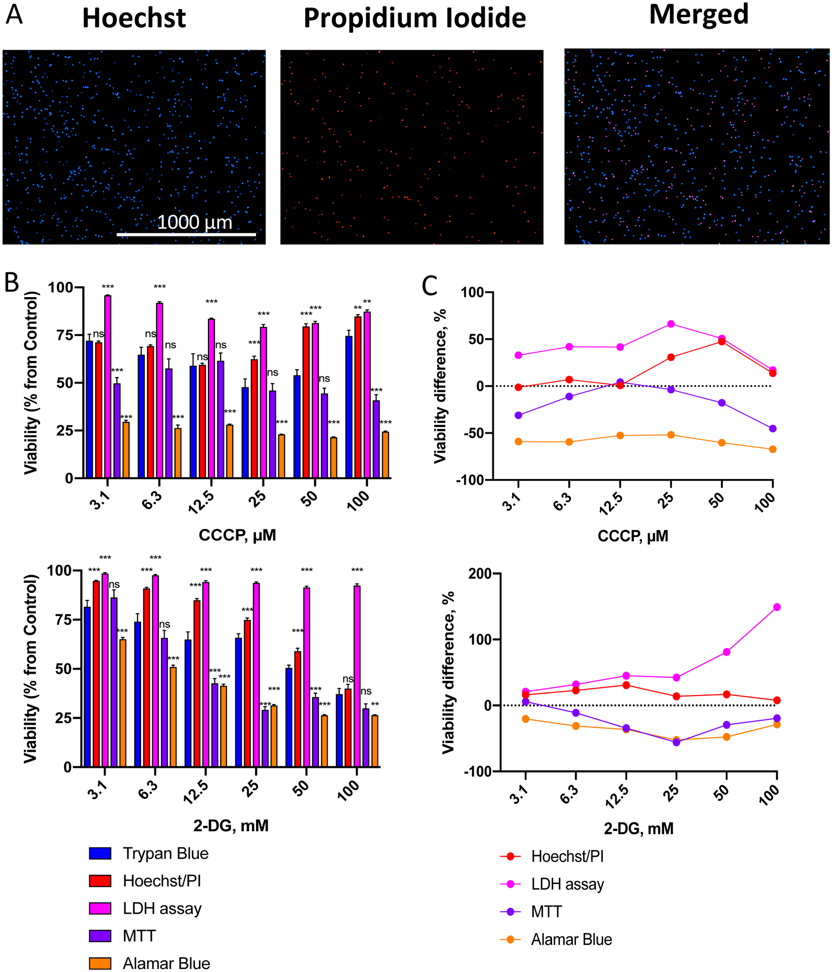
Figure 2. Comparison of a panel of cytotoxicity assays with trypan blue exclusion.

A. Representative images of total (Hoechst 33342) and dead (propidium iodide, PI) OCI-AML2 cells via Hoechst/PI staining. B. Assessment of viability of OCI-AML2 cells using different methods after treatment with a gradient of CCCP (top) or 2-DG (bottom) concentrations. OCI-AML2 were treated with CCCP or 2-DG in serum-free RPMI-1640 for 24 h prior to determination of cell viability. Shown is mean, error bars represent SEM. C. Difference in cell viability between cytotoxicity assays in (B) vs. trypan blue staining (see Supplementary Tables S1–2 for the exact numbers and median difference). Stars indicate significant difference vs. trypan blue staining. ** p < 0.01, *** p < 0.001, ns – non-significant. Group comparison was done via t-test with correction for multiple hypothesis testing. Three independent biological replicates were performed.
