Fig. 1.
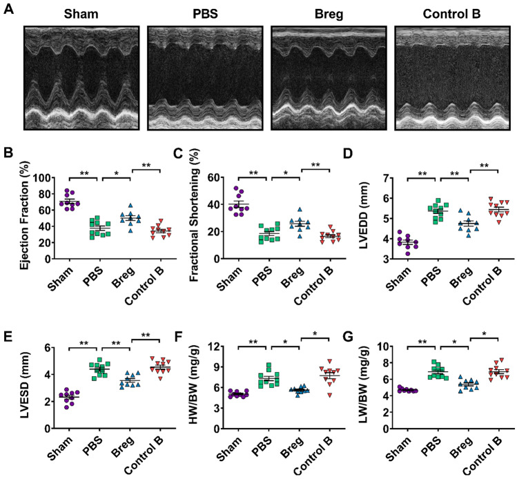
Adoptive transfer of Bregs improves cardiac function after MI. a Representative M‐mode echocardiographic images of the left ventricle 28 days after MI. b–e Analysis of ejection fraction (b), fractional shortening (c), LVEDD (d) and LVESD (e) by echocardiography at day 28 after MI. n = 9–10 per group. f, g HW/BW and LW/HW were measured at day 28 after MI. n = 9–10 per group. Data are expressed as means ± SEM. *P < 0.05, **P < 0.01. Data in b–e were analyzed by one-way ANOVA, followed by Tukey’s post hoc test. Data in f and g were analyzed by Kruskal–Wallis test with Dunn’s multiple comparisons test. Sham sham-operated group, PBS MI mice that received phosphate buffered saline, Breg MI mice that received regulatory B cells, Control B MI mice that received control B cells, LVEDD left ventricular end-diastolic dimension, LVESD left ventricular end-systolic dimension, HW/BW heart weight/body weight ratio, LW/BW lung weight/body weight ratio

