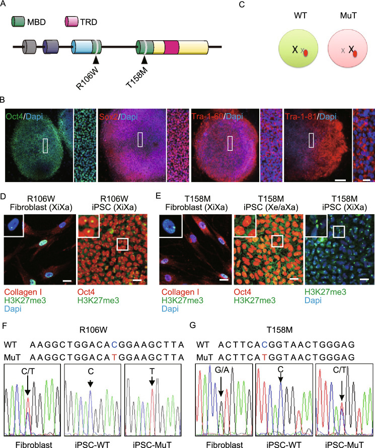
Figure 1.
Generation of iPSCs from RTT patient dermal fibroblasts. (A) A schematic representing MeCP2 gene structure showing the two different point mutations analyzed in this study. MBD: methyl-CpG binding domain; TRD: transcription repression domain. (B) Representative images of RTT-iPSCs showing expression of pluripotency markers Oct4, Sox2, Tra-1-81 and Tra-1-60 as well as typical hESC colony-like morphology (Bars: 250 μm and 50 μm). (C) A schematic representing X inactivation. In the brain of RTT patients, MeCP2 mutation is chimeric due to random X inactivation. (D) Representative images of H3K27me3 immunofluorescence in RTT fibroblasts and iPSCs. All fibroblast lines exhibit predominant H3K27me3 nuclear foci (Barr body) and express fibroblast marker, collagen I. R106W iPSCs also show clear H3K27me3 foci and pluripotent marker Oct4. (E) T158M (Xa/Xa or Xa/Xe) iPSCs show diffused immune reactivity indicating the absence of Barr body (Bar: 25 μm). (F) Mono-allelic expression of MeCP2 in R106W hiPSC lines. Sequencing of MeCP2 cDNA from R106W fibroblasts showing expression of both WT and mutant MeCP2, whereas R106W iPSCs only express one of the two alleles. This is likely due to the retention of the XCI status of the individual fibroblast from which each of these hiPSC lines originates. (G) MeCP2 cDNA sequencing results from T158M fibroblasts and iPSCs showing expression of both WT and MuT MeCP2 alleles in multi clonal fibroblast cultures and in one heterogeneous (XaXe or Xa/Xa) T158M iPSC line, whereas other iPSC lines only express the WT allele

