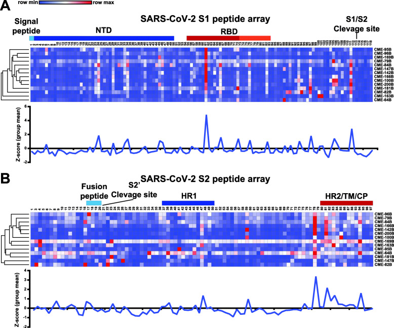
Fig. 5.
High-resolution mapping of SARS-CoV-2 antibody responses after COVID-19 vaccine. A, B Plasma IgG binding to SARS-CoV-2 peptides spanning the SARS-CoV-2 spike protein (A S1 and B S2 subunits separated;12-mer overlapping peptides) in 14 SARS-CoV-2 seronegative adults after primary COVID-19 immunization (week 3). Each peptide was printed in triplicate and the log2 of the average median fluorescent intensity (F635) for each peptide graphed. Row color corresponds to the minimum and maximum intensity for all peptides for each individual. Known regions of the spike protein are annotated above heatmaps. Orange section in RBD depicts critical ACE2 contact residues. Line graphs displaying the mean group Z-score for each peptide in S1 and S2 from the vaccine group (blue; n = 14)

