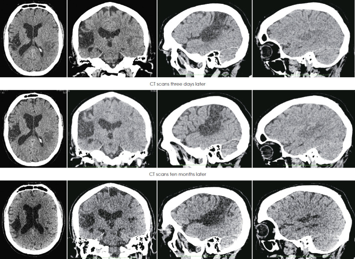
Fig. 3.
CT of the brain performed on the day of the ischaemic stroke (first row), three days later (second row) and ten months later (third row). The scans show former massive ischaemic infarct lesion in the right hemisphere and evolution of the smaller acute ischemic infarct in the left temporal lobe. The first column presents axial section scans, second column coronal, third sagittal of the right hemisphere, and fourth sagittal of the left hemisphere.

