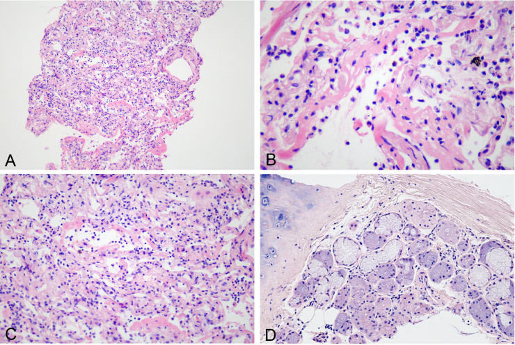
Figure 1.
Histological changes of exudative phase of COVID-19 pneumonia seen in patient 1. (A) Focal alveolar collapse (H&E 10×). (B) Hyaline membrane formation (H&E 40×). (C) Widened alveolar septa and lymphocytic infiltration (H&E 20×). (D) Bronchial submucosal glands with a few lymphocytes in the stroma (H&E 20×).

