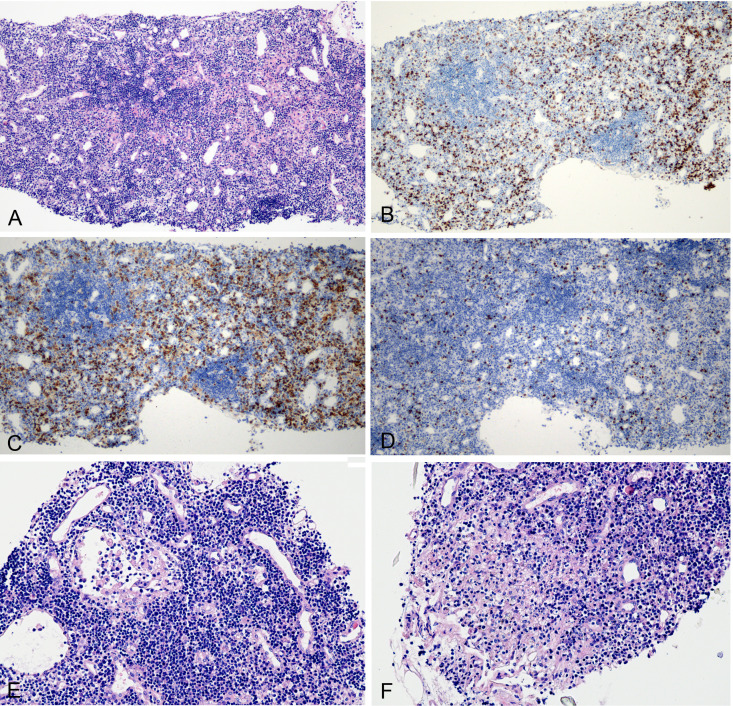
Figure 3.
Histological changes of intrathoracic lymph node. (A) Primary lymphoid follicles with slightly widened interfollicular zone (H&E 10×). (B) CD3-positive lymphocytes (immunohistochemical (IHC) stain 10×). (C) CD4-positive stain lymphocytes (IHC stain, 10×). (D) CD8-positive stain lymphocytes (IHC stain, 10×). (E) Lymphatic sinus dilation and vascular endothelial hyperplasia (H&E 20×). (F) Focal necrosis (H&E 20×).

