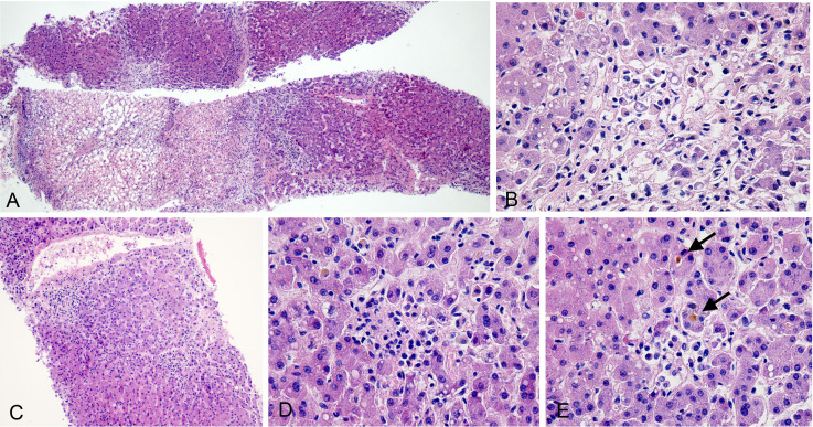
Figure 5.
Histological changes of the liver. (A) Large areas of coagulative necrosis (H&E 4×). (B) Focal coagulative necrosis without obvious inflammation (H&E 40×). (C) Inflammation around a central vein (H&E 10×). (D) Small focus of confluent necrosis with mild mononuclear cell infiltration (H&E 40×). (E) Canalicular cholestasis (arrow; H&E 40×).

