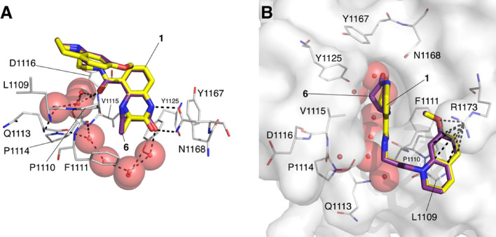
Figure 3.
X-ray crystal structure of 6 bound to the CREBBP bromodomain (PDB code: 6YIM; carbon = purple; protein surface from this structure shown) overlaid with the X-ray crystal structure of 1 bound to the CREBBP bromodomain (PDB code: 4NYX; carbon = yellow).11 (A) The side orientation shows that the headgroups of each compound form the same hydrogen-bonding interactions with the bromodomain and that the KAc-mimicking methyl and carbonyl groups of both molecules overlay very closely. (B) The top orientation shows that the interaction with R1173 is present for both molecules. The kink in the 4,5-dihydrobenzodiazepinone ring of 6 (carbon = purple), which more fully occupies the KAc-binding pocket, is visible. See Figure S10 for the ligand electron density map.

