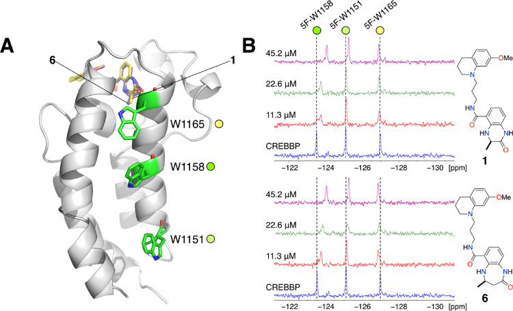
Figure 4.
(A) X-ray crystal structure of 6 bound to the CREBBP bromodomain (PDB code: 6YIM; carbon = purple; cartoon from this structure shown) overlaid with the X-ray crystal structure of 1 bound to the CREBBP bromodomain (PDB code: 4NYX; carbon = yellow).11 The three tryptophan residues (W1151, W1158, and W1165) that were replaced by 5-fluorotryptophan are highlighted as green sticks. (B) Partial 19F NMR spectra showing the effect of adding 11.3, 22.6, or 45.2 μM of either 1 or 6 to the 5-fluorotryptophan-containing CREBBP bromodomain (45 μM). The resonances were assigned to specific 5-FW residues using point mutation studies (Figure S4C).

