
Figure 2.
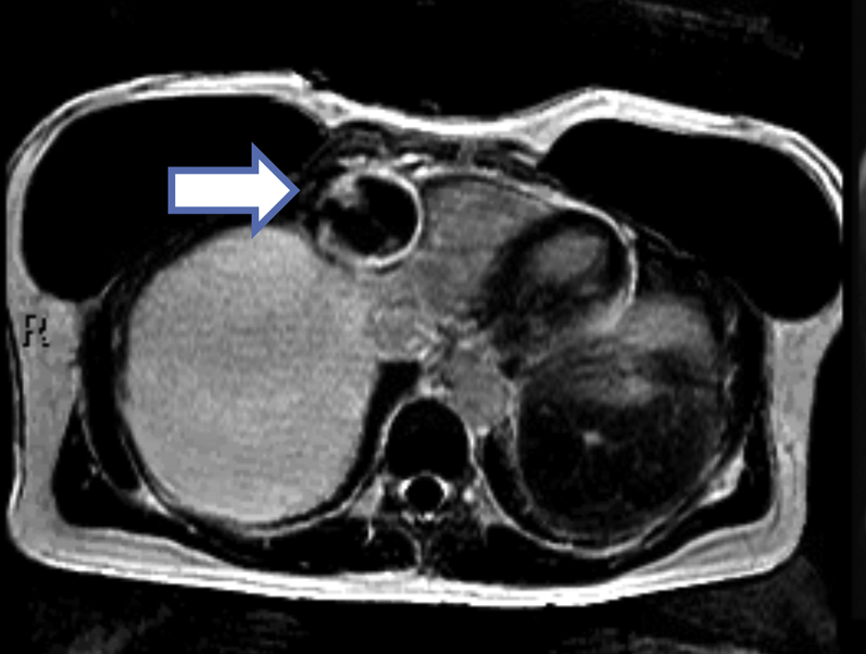
Cardiac Magnetic Resonance Imaging
Axial slice cardiac magnetic resonance with a phase-sensitive inversion recovery sequence showing partial filling of the aneurysm along with a rim of delayed gadolinium enhancement in the periphery of the aneurysm (arrow).