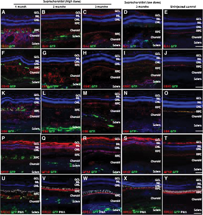
Figure 2.
Local immune cell infiltration after suprachoroidal delivery of AAV8 in NHP eyes. (A–Y) Confocal fluorescence images of GFP transgene expression (green) co-immunostained with antibodies to IBA-1+ microglial cells (A–E), CD45+ leukocytes (F–J), CD8+ cytotoxic T cells (K–O), and GFAP+ reactive gliosis (P–T), as well as RPE65 (red) to label RPE cells and PNA (white) to label cone photoreceptor inner/out segments, along with DAPI (blue) to label cell nuclei in eyes at 1 month (A, F, K, P, and U), 2 months (B, G, L, Q, and V), and 3 months (C, H, M, R, and W) after high-dose or low-dose (D, I, N, S, and X) suprachoroidal AAV8 injections, compared to uninjected control eyes (E, J, O, T, and V). Scale bars: 100 μm. GCL, ganglion cell layers; GFAP, glial fibrillary acidic protein; IBA-1, ionized calcium-binding adaptor-1; INL, inner nuclear layer; ONL, outer nuclear layer; PNA, peanut agglutinin; RPE, retinal pigment epithelium. Color images are available online.

