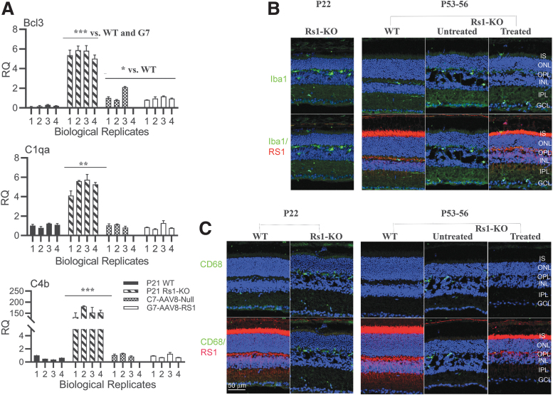
Figure 4.
AAV8-RS1 attenuates inflammatory responses and restores immune quiescence state in Rs1-KO (Rs1−/y) retina. (A) Exogenous RS1 gene expression in Rs1-KO (Rs1-/y) retina suppresses pro- inflammatory C1qa, C4b, andBcl3 gene expression. Bar graphs of RNA expression for indicated genes in WT, Rs1-KO, C7 and G7 retinas. Experiment is described in Fig 2A. Data are expressed as Mean ± SEM. Statistical significance determined using the Holm-Sidak method, with alpha = 0.05. Each row was analyzed individually, without assuming a consistent SD. *p ≤ 0.05, **p ≤ 0.01, ***p ≤ 0.001. (B) Exogenous RS1 gene expression inhibits microglial activation in Rs1-KO retina: Retinal sections double stained Iba (1:500, green) and Rs1 (1:1000, red). Microglia in age matched WT retinas showed a tiny cell soma, little perinuclear cytoplasm, and a small number of fine, branched processes covered in numerous projections. In the untreated Rs1-KO retinas at P21 and P49 an increase in microglial cell number was found compared with WT control retinas (Figure 1B). Moreover, numerous amoeboid Iba1 positive cells were observed in all retinal layers, with greater presence in IPL and the OPL. In retinas expressing exogenous RS1 gene amoeboid Iba1 -positive cells were less abundant than in untreated in all layers of the retina and were mainly distributed in more internal GC layer. (C) Retinal sections double stained for RS1(1:1000, red) and CD68 (1:400, green). CD68 is a phagocytic marker associated with the involvement of monocytes/macrophages. CD68 staining is undetectable in age matched WT retinas and CD68 staining is intense in OPL in untreated RS1-KO retina. CD68 staining barely detectable in treated retina. Data from one representative experiment of two or three independent experiments. Scale bars: 50 μm. n = 3.

