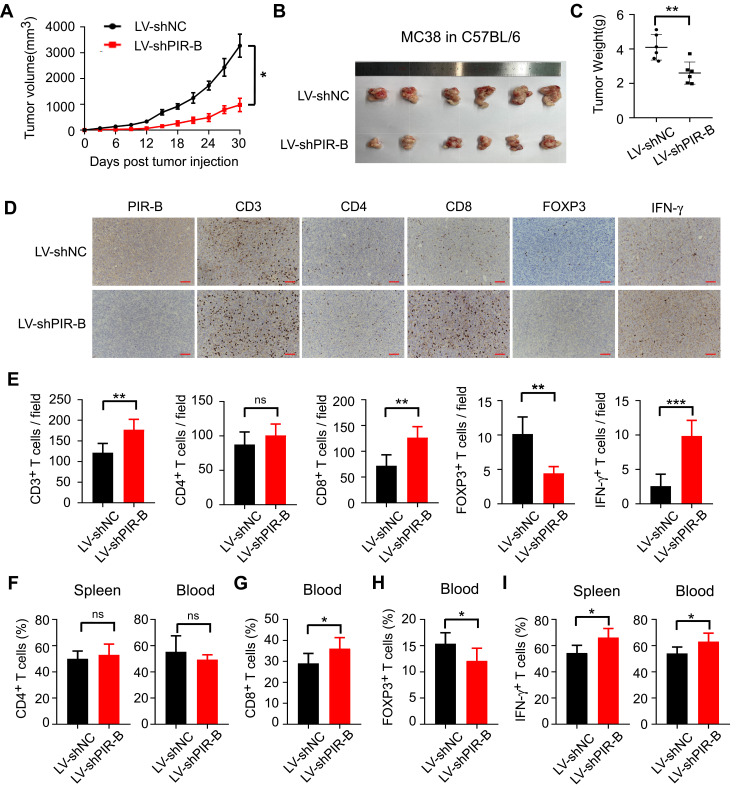
Figure 7.
PIR-B knockdown inhibited immunosuppressive T cell contexture and tumor progression in vivo. (A) PIR-B knockdown profoundly slowed MC38 tumor growth in C57BL/6 mice. MC38 cells (1×10⁷ cells/mouse) infected with PIR-B knockdown or control lentivirus were subcutaneously inoculated into 6–8-week female C57BL/6 mice. Tumor sizes were measured every three days and are presented as mean ± SD (n = 6 mice/group). (B and C) PIR-B knockdown in MC38 cells markedly decreased final tumor sizes and weights of transplanted tumors. (B) Tumor sizes and (C) mean ± SD of tumor weights from each group at the endpoint of the experiments. (D and E) Tumor tissues with PIR-B knockdown displayed markedly higher CD3+, CD8+, and IFN-γ+ T cell densities, but lower FOXP3+ T cell densities than those in the control group. The numbers of T cell subsets were determined by IHC analysis. (D) Representative images of IHC analysis. (E) Mean ± SD of different T cell subset infiltration in five random fields. (F) Flow cytometric analysis showed that PIR-B knockdown did not affect CD4+ T cell frequency in mouse blood and spleens. (G and H) Flow cytometric analysis showed that PIR-B knockdown remarkably increased the frequency of CD8+ T cells but decreased that of FOXP3+ Tregs. (G) The fraction of CD8+ T cells among CD3+ T cells. (H) The fraction of FOXP3+ T cells among CD3+ T cells. (I) Flow cytometric analysis showed that the T cells from the spleens and blood of PIR-B knockdown mice produced markedly more IFN-γ than the spleens and blood of the control group mice. The total T cells were calculated from the CD3+ cells in both organs. *p<0.05; **p<0.01; ***p<0.001.
Abbreviations: LV-shNC, MC38 cells transfected with lentiviruses carrying control shRNA; LV-shPIR-B, MC38 cells transfected with lentiviruses carrying specific PIR-B shRNA.

