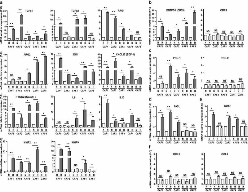
Figure 4.

Hypoxic conditioning of CAFs improves their expression of several immune-suppressive factors. RT-qPCR analysis of (a) TGF-β1, TGF-β2 ARG1, ARG2, IDO1, CXCL12 (SDF-1), PTGS2 (Cox2), IL6, IL10, MMP2, MMP9, (b) ENTPD1 (CD39), CD73, (c) PD-L1, PD-L2, (d) FasL, (e) CD47, (f) CCL2 and CCL5 expression by hypoxic versus normoxic CAFs. Results are expressed as the mean ± s.d. of three independent experiments, normalized to “1” in normoxic conditions (N: Normoxia; H: Hypoxia). P values were determined by unpaired two-tailed student’s t-test. (NS: non-significant; *p < .05; ** p < .01)
