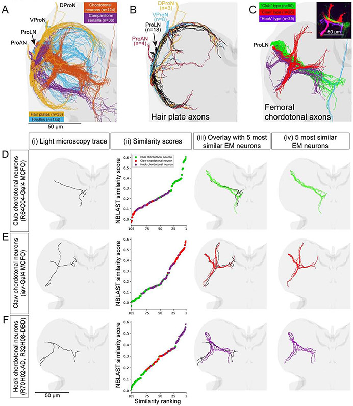
Figure 4. Identification of sensory neuron subtypes.
(A) Reconstruction of the main branches of sensory axons for the front left leg. The four main functional subtypes of sensory neurons (different colors) are identifiable from their projection patterns. Light grey, VNC. Darker grey, neuropil. ProAN, prothoracic accessory nerve; ProLN, prothoracic leg nerve; VProN, ventral prothoracic nerve; DProN, dorsal prothoracic nerve.
(B) Organization of hair plate neuron projections. Hair plate axons enter the T1 neuromere through four different nerves (different colors) and branch to encircle the neuromere.
(C) Femoral chordotonal organ (FeCO) neuron subtypes. Inset: Different subtypes, characterized previously with light microscopy (LM), encode different aspects of leg kinematics (adapted from Mamiya et al., 2018).
(D-E) Comparison of EM reconstructions with LM reconstructions from genetic driver lines that specifically label FeCO neurons (Mamiya et al. 2018, Chen et al. in preparation). (i) Rendering of LM reconstruction. (ii) Ranked distribution of NBLAST similarity scores (worst to best, left to right) color coded by FeCO neuron subtype (as in C). (iii) Overlay of the LM reconstruction and the five most similar EM reconstructions. (iv) The five most similar EM reconstructions alone.
(D) A club FeCO neuron (MCFO from R64C04-Gal4).
(E) A claw FeCO neuron (MCFO from iav-Gal4).
(F) A hook FeCO neuron (R70H02-AD, R32H08-DBD).
Scale bars, 50 μm (A-F).

