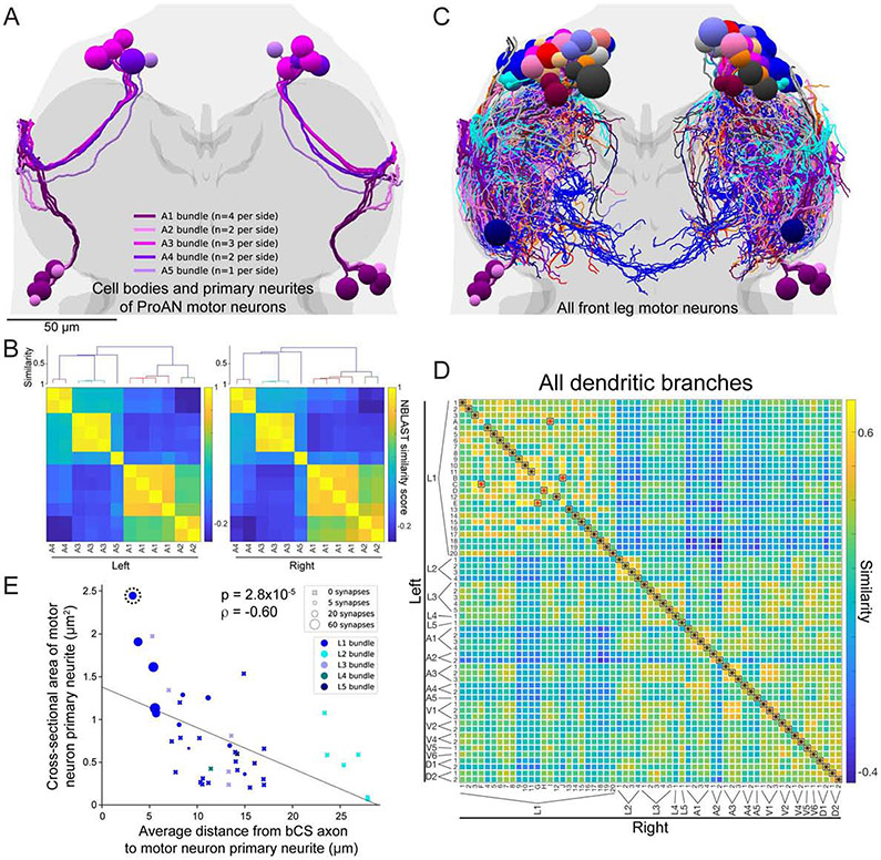
Figure 6. MN bundles, symmetry, uniqueness, and bCS connectivity.
(A) Reconstruction of cell bodies and primary neurites of the 24 ProAN MNs (12 per side). Primary neurites travel through the neuromere in five distinct and highly symmetric bundles (numbered A1 through A5, colored in shades of purple). See also Fig. S6.
(B) Quantitative analysis of bundles of MN primary neurites. ProAN MNs on each side of the VNC were clustered by the similarity in primary neurite positions (STAR Methods). Top, dendrogram from hierarchical clustering. Members of each bundle cluster together. Bottom, matrix of NBLAST similarity scores.
(C) Branching patterns of all 139 MNs arborizing in the T1 neuromeres were reconstructed and transformed into the atlas coordinate system (Fig. S3).
(D) Identification of left–right homologous pairs of front leg MNs. Of the 69 left and 70 right T1 MNs, expert annotators identified 61 symmetric left–right pairs. A global pairwise assignment of NBLAST similarity scores agreed on 92% (56 of 61) of identified pairs. Black asterisks, agreements. Red asterisks, disagreements.
(E) Relationship between four anatomical properties of leg MNs: proximity to bCS axons (x-axis), primary neurite cross-sectional area (y-axis), primary neurite bundle (marker color), and number of synapses received from bCS neurons (marker type and size). MNs closer to bCS axons have larger-caliber primary neurites. Only MNs in the L1 bundle received any synapses from bCS neurons. Within the L1 bundle, those receiving the most synapses have large-caliber primary neurites and are closest to bCS axons. Dashed circle indicates the MN whose morphology is most similar to a functionally characterized fast flexor MN (Fig. 7A).
Scale bars, 50 μm (A, C).

