Figure 7. A fast tibia flexor motor neuron is a major synaptic target of bCS neurons.
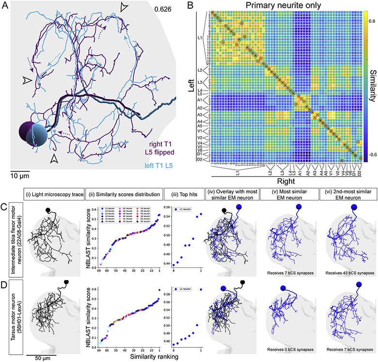
(A-C) MNs reconstructed from LM matched to the most similar neurons reconstructed from EM. (i) Rendering of LM reconstruction. (ii) Ranked distribution of NBLAST similarity scores (worst to best, left to right) color coded by MN bundle (key, Aii top). (iii) Zoom-in on the 8 highest similarity scores. (iv) Overlay of the LM reconstruction and the most similar EM reconstruction. (v) The most similar EM reconstruction. (vi) The second-most similar EM reconstruction.
(A) A fast tibia flexor MN (81A07-Gal4). The two most similar EM reconstructions both receive strong synaptic input from the two left and two right T1 bCS neurons.
(B) A slow tibia flexor MN (35C09-Gal4). The two most similar EM reconstructions receive minimal synaptic input from T1 bCS neurons.
(C) A MN innervating the tibia long tendon muscle, which controls movements of the tarsus (21G01-LexA). The two most similar EM reconstructions receive no synaptic input from T1 bCS neurons.
Scale bars, 50 μm (A-C).

