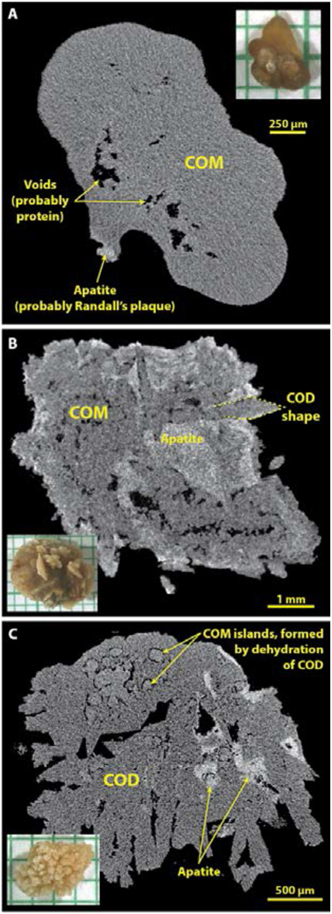
Figure 4.
Typical morphologies of calcium oxalate stones as seen by micro CT. Insets show photographs of stones on mm paper. A: Type Ia stone, probably on Randall’s plaque, shows the rather homogeneous gray appearance of tightly packed COM. B: Stone showing COD crystal shapes but in which the COD has been almost entirely converted to COM. C: Stone composed mainly of COD, but showing the beginning stages of conversion to COM. In stones forming as COD crystals, it was common to see the interstices filled with apatite (panels B and C), as has been previously described.[30]

