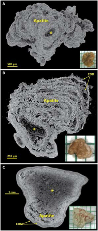
Figure 5.
Apatite stones most commonly show alternation of X-ray-dense and more lucent layers, and often relatively large X-ray-lucent regions (*). Spectroscopic analysis of X-ray-lucent regions in apatite stones has always shown only apatite, but sometimes with an increase in the protein content of the material. A: A typical apatite stone, pure by spectroscopic analysis. B: An example of the IVa+IIa morphology, with COD crystals between apatite layers. C: An apatite stone that has thin layers of COM at its surface.

