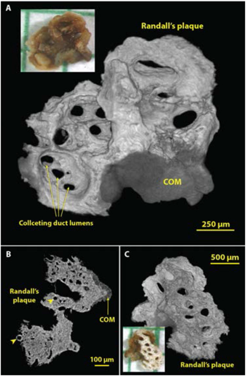
Figure 9.
Using micro CT to visualize structures in three dimensions (3D). Shown are images of a small calculus that had been adherent to the renal papilla. The bulk of the ‘stone’ was actually well-developed Randall’s plaque, with a small region of COM overgrowth from the urine. A: 3D surface rendering showing the side of the calculus that faced the urine. Lumens of collecting ducts (about 100-150 μm in diameter) through the mineralized plaque are obvious. B: Micro CT slice through part of the calculus showing COM overgrowth and the lumens of thin limbs and vessels within the plaque region (arrowheads). C: Surface rendering of the underside of the calculus. Micro CT scan of this calculus was high resolution (2.08 μm voxel size).

