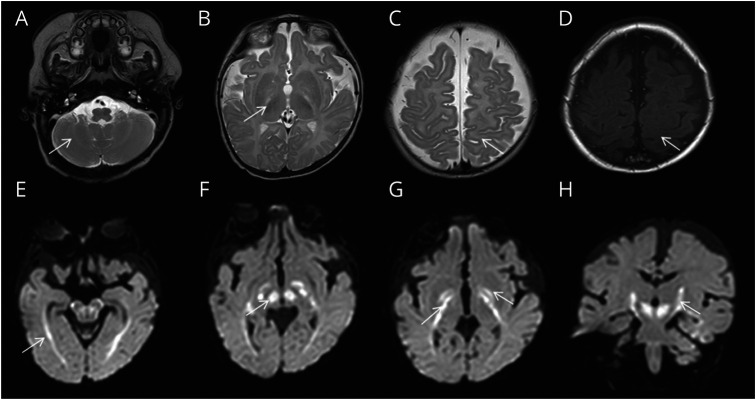
Figure. MRI Brain at Age 4 Months.
T2 (A–C) and T1-weighted (D) images depict lack of myelination in cerebellar white matter, posterior limb of internal capsule, and perirolandic regions (arrows). Diffusion-weighted imaging (E–H) shows diffusion restriction involving optic radiations (E), red nucleus and cerebral peduncle region (F), globus pallidus, and along corticospinal tract (G and H).

