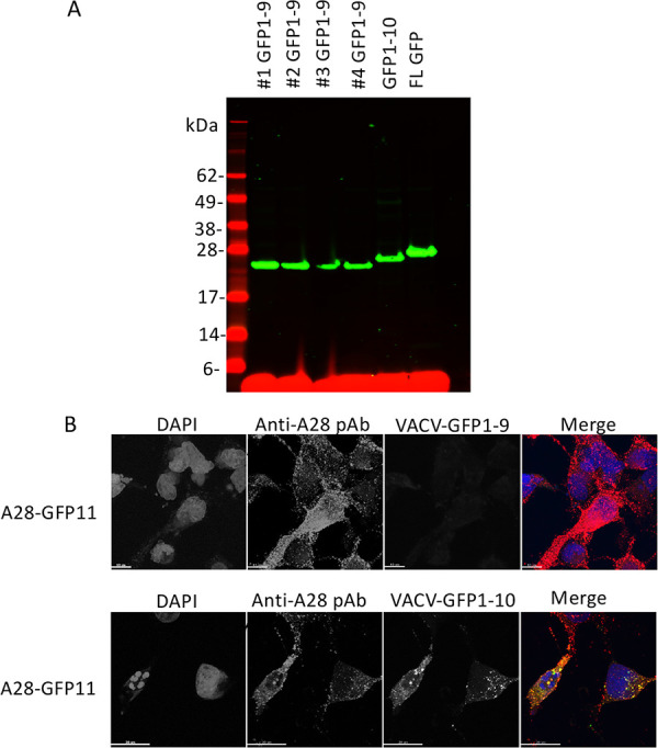
FIG 3.

Expression of recombinant GFP1-9 and GFP1-10 by recombinant VACV. (A) RK13 cells were infected with four individual clones of VACV-GFP1-9, VACV-GFP1-10, and a VACV expressing full-length (FL) GFP. Lysates were analyzed by SDS-PAGE, followed by Western blotting with an anti-GFP antibody and a secondary fluorescent antibody. Molecular weight markers are shown on the left. (B) RK13 cells on coverslips were infected with VACV-GFP1-9 or VACV-GFP1-10 and transfected with a plasmid expressing A28-V5-GFP11. After overnight incubation at 37°C, the cells were fixed, permeabilized, and stained with polyclonal antibody (pAb) to A28, followed by fluorescent secondary antibody and DAPI. Green fluorescence was detected in cells infected with VACV-GFP1-10 but not with VACV-GFP1-9. The merge shows DAPI (blue), anti-A28 (red), GFP (green), and overlap of anti-A28 and GFP (yellow).
