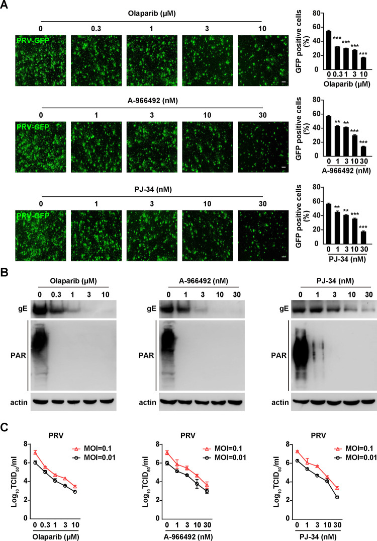
FIG 2.
PARP1 inhibitors exhibit inhibitory effect on PRV infection. (A) PK15 cells were infected with PRV-GFP (MOI = 0.01) and treated with olaparib (0 to 10 μM), A-966492 (0 to 30 nM), or PJ-34 (0 to 30 nM) for 36 h. Viral replication was analyzed by fluorescence microscopy, and GFP-positive cells were analyzed by flow cytometry. Bars, 100 μm. (B) PK15 cells were infected with PRV-QXX (MOI = 0.1) and treated with olaparib (0 to 10 μM), A-966492 (0 to 30 nM), or PJ-34 (0 to 30 nM) for 24 h. PRV gE, PAR, and actin were assessed by immunoblotting analysis. (C) PK15 cells were infected with PRV-QXX (MOI = 0.01 and 0.1) and treated with olaparib (0 to 10 μM), A-966492 (0 to 30 nM), or PJ-34 (0 to 30 nM) for 24 h. PRV titers were assessed by TCID50 assay. Data are means and SD based on three independent experiments. **, P < 0.01, and ***, P < 0.001, determined by two-tailed Student's t test.

