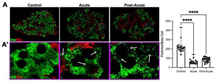
Figure 3.
Beta cells from SARS-COV-2 subjects are significantly degranulated. (A) Immunohistochemical staining for insulin and glucagon of representative islets that were imaged using super-resolution microscopy. (A’) High-magnification images purple boxed areas in (A). White arrows highlight large areas in the cytoplasm with reduced insulin staining in pancreas from SCV-inoculated subjects. (B) Quantification of beta cell granulation revealed a greater than 60% decrease in granulation between control and acute/post-acute islets (n=10–20 islets per group from at least 3 different subjects. Each dot represents 1 islet. Two-way ANOVA with Turkey’s multiple comparisons, **** p<0.001)

