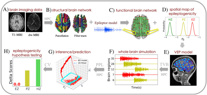
Fig 1. The workflow of BVEP aims at assessing different hypotheses regarding the location of an epileptogenic zone in a personalized whole-brain model of epilepsy spread.
(A) At the first step, the patient undergoes non-invasive brain imaging such as MRI, and diffusion-weighted MRI (dwMRI). (B) Based on these images, the structural brain network model including brain parcellation and fiber tracts of the human brain are generated. (C) Then, a neural mass model (here 6D Epileptor) is defined at each brain region and connected through the structural connectivity matrix to build the functional brain network model. (D, E) Next, the VEP brain model is constructed by furnishing the functional brain network model with the spatial map of epileptogenicity (EZ, PZ, HZ hypotheses) across different brain regions. (F) The simulations by TVB allow to specifically mimic the individual’s spatiotemporal macro-level brain activity. (G) Subsequently, the generative VEP brain model is embedded within a PPL tool to invert the BVEP brain model (here 2D Epileptor) and evaluating the model against the patient’s data. (H) Finally, cross-validation (CV) is performed using WAIC/LOO to assess the model’s prediction performance on new data, in order to evaluate and validate the quality of the hypotheses regarding epileptogenicity.

