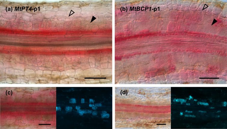
Fig 3. Betalain accumulation is observed at different intensities in different tissue layers and appears higher in the endodermal cells adjacent to arbuscule-containing cortical cells.
Root sections of Medicago truncatula expressing MtPT4-p1 (a and c) and MtBCP1-p1 (b and d) 4 weeks after inoculation with Rhizophagus irregularis. (c and d) Left: Betacyanin pigments are visible in red; right: WGA-FITC staining of fungal structures in blue. Open arrows mark internal hyphae, and filled arrows signal cells containing arbuscules. Scale bar, 100 μm. WGA, wheat germ agglutinin.

