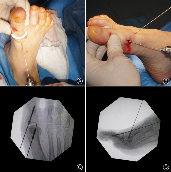
Fig. 2.

(A): Clinical photo showing the correction maneuver of the deformity (force applied as demonstrated with arrow). (B): Applying hollow nail guidewires for fixation of the fragment. Radiography showing placement of the screw percutaneously after osteotomy. (C): A‐P view; (D): lateral view.
