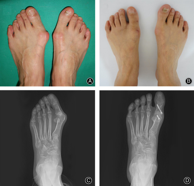
Fig. 4.

Clinical and radiographic view of a 60‐year‐old female with a left hallux valgus. Preoperative clinical photo: preoperative (A) and postoperative 24 months follow‐up (B); radiographic X‐ray: preoperative (C) and postoperative 24 months follow‐up (D).
