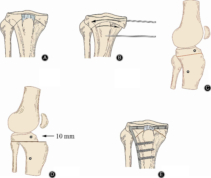
Fig 3.

The method dome‐shaped hightibial osteotomy. (A) Series of 2.5‐mm drill holes marked on a curved line above the tibial tuberosity. (B) Two Steinmann pins inserted on either side of the osteotomy to define angular correction. (C/D) Sagittal view: the distal tibia brought forward approximately 10 mm. (E) When desired angle is achieved, the TomoFix plate fix fragments are applied under compression. The figure was adapted from Diogo et al. 47
