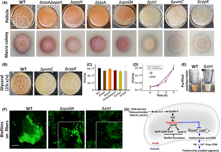
Fig. 2.

Biofilm formation and pulcherrimin production by B. subtilis (WT and mutants cells) grown in chickpea milk.
A. Pellicle, pigment and colony‐type biofilm formations by the WT and mutants cells in chickpea milk (CPM) or CPM agar after 72 h of incubation at 30°C.
B. Pellicle formations by pulcherrimin‐deficient mutants in CPM in the presence or absence of glycerol (1% v/v) after 72 h of incubation at 30°C.
C. Colony forming units (CFUs) of the WT andmutant cells in CPM after 24 h of incubation at 37°C. The graph shows the means ± SEMs of three measurements. *P < 0.05 vs. the non‐treated controls.
D. Growth curve of B. subtilis WT cells in LB and CPM grown at 37°C with 150 rpm shaking. The graph shows the means ± SEMs of three measurements.
E. Purified pulcherrimin (in methanol) from WT, and sinI mutants. Detailed processing method is depicted in Fig. S5.
F. Interactions of B. subtilis WT, spo0A and sinI mutants (displaying intense green fluorescence) with CPM fibres (faint green autofluorescence).
G. A schematic representation depicting the regulation of biofilm formation and pulcherrimin production pathways in B. subtilis grown in CPM.
