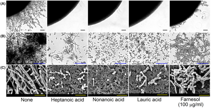
Fig. 2.

Inhibition of hyphal filamentation and aggregation by medium‐chain fatty acids. C. albicans morphology on solid media (A). C. albicans was streaked on PDA solid plates in the absence or presence of heptanoic acid (7:0), nonanoic acid (9:0) or lauric acid (12:0) at 2 µg ml−1 or farnesol at 100 µg ml−1. Colony morphologies were observed during incubation for 6 days at 37°C. Inhibition of filamentation and of cell aggregation in PDB medium (B). Hyphae were visualized after incubation for 24 h. The scale bars in panels A and B represent 100 µm. None; non‐treated control. SEM observation of hyphal inhibition in C. albicans biofilms grown in PDB medium by fatty acids (C). The scale bars represent 15 µm.
