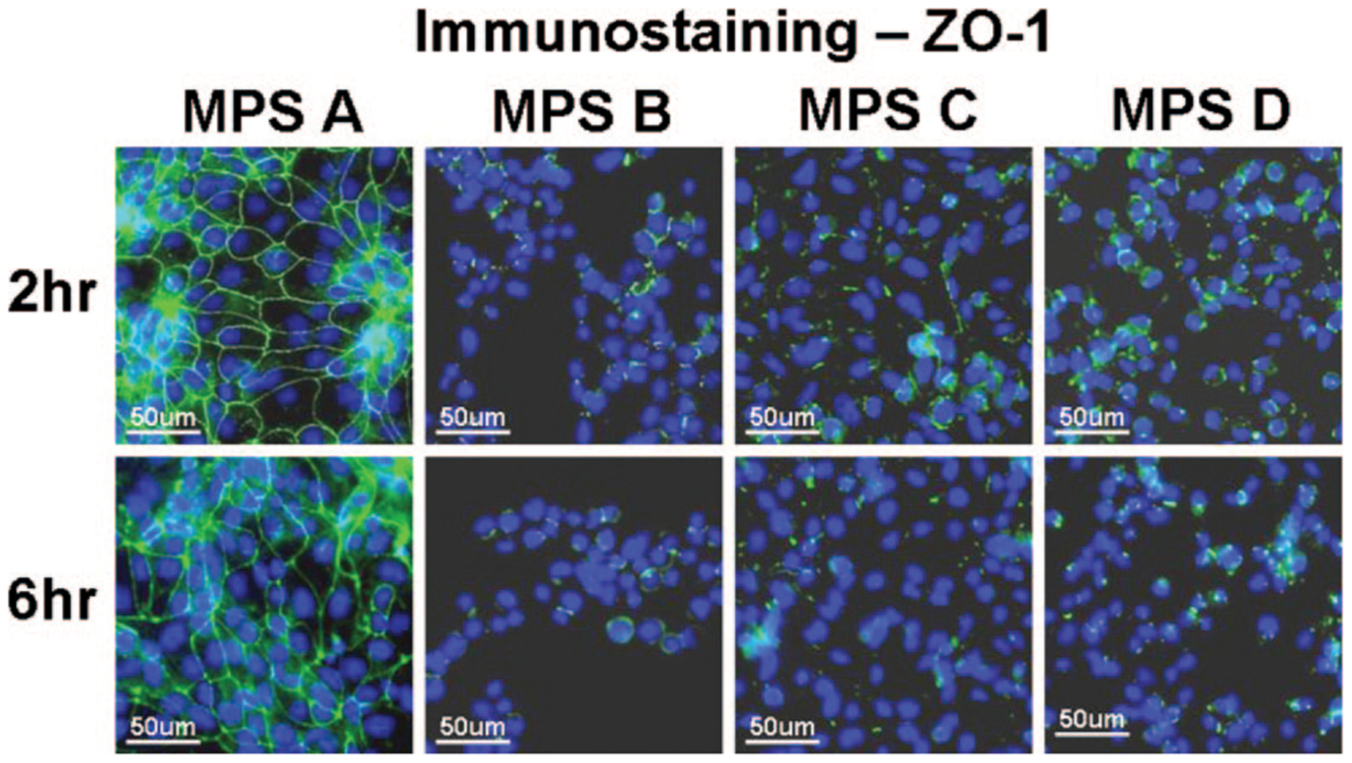
FIG. 5.

Representative images of immunofluorescent staining for ZO-1 (green) in human corneal epithelial cells after 2 and 6 hrs exposure to each of the 4 MPS studied. Strong green staining between the cells indicates intact ZO-1 tight junction proteins while the weak or negative staining shows disrupted junctions. Hoechst 33342 (blue) was used for cell nuclear counterstaining.
