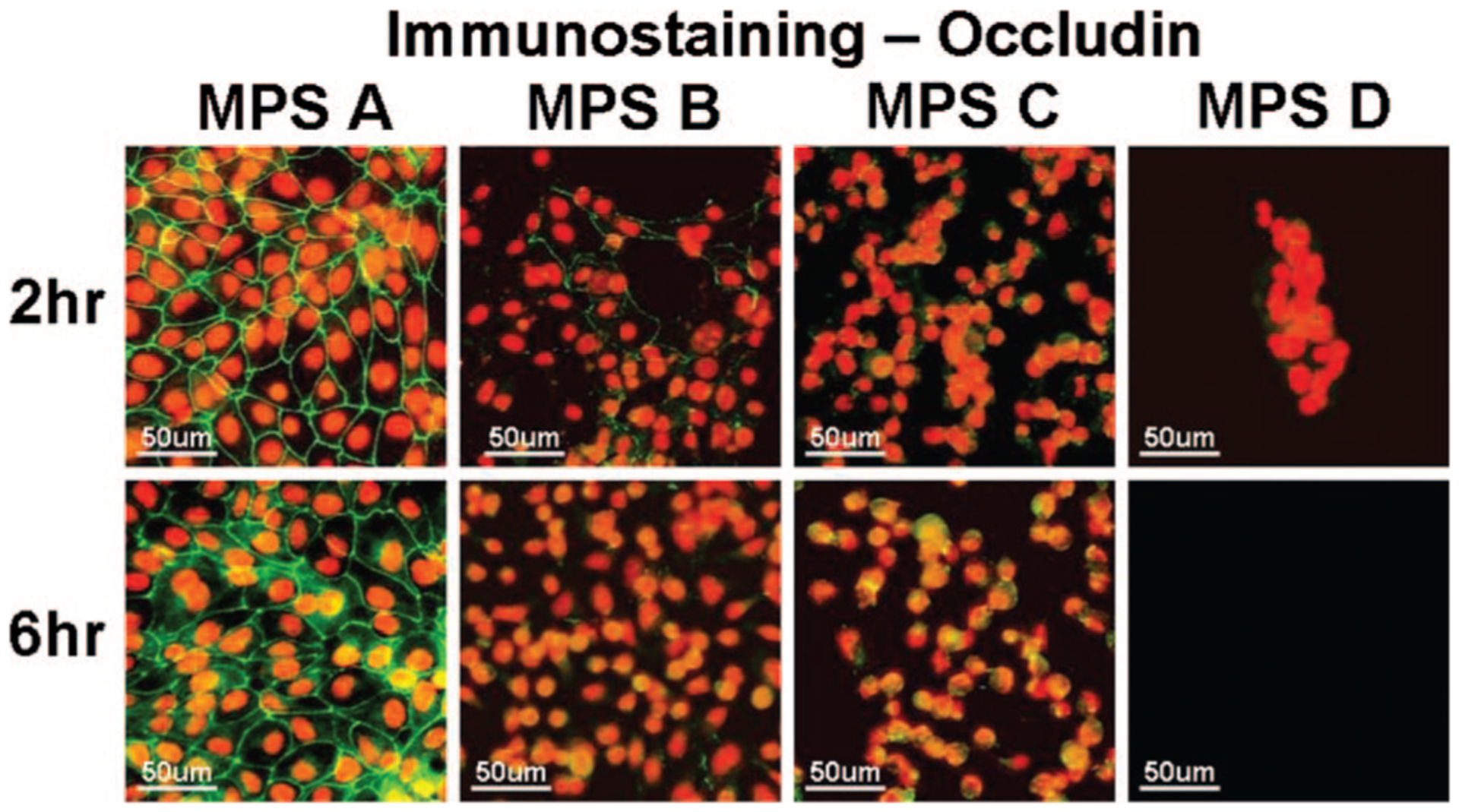
FIG. 6.

Representative images of immunofluorescent staining for occludin (green) in human corneal epithelial cells after 2 and 6 hrs exposure to each of the 4 MPS studied. Strong green staining between the cells indicates intact occludin tight junction proteins while the weak or negative staining shows disrupted junctions. PI (red) was used for cell nuclear counterstaining.
