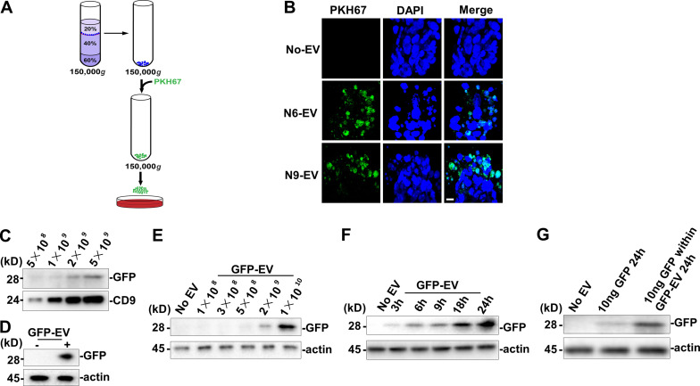
Figure 3.
Differentiated neuronal EVs taken up by mESCs.(A) Schematic of mESCs treated by PKH6 dye–labeled EVs. (B) Confocal images of differentiated mESCs incubated without EV (No-EV) or with PKH6 dye–labeled N6 or N9 EV. Nuclei were stained with DAPI. Scale bar, 10 µm. (C) Immunoblot of GFP and CD9 in EVs (GFP-EV) purified from GFP-expressing cells. 2 × 10⁵ mESCs in a 35-mm dish were incubated in 2 ml of N2B27 medium for 24 h with EVs purified from control or GFP-expressing cells. (D) Immunoblot of GFP from whole-cell lysate of mESCs treated with PBS or GFP-EV for 24 h. (E) Immunoblot of GFP from whole-cell lysate of mESCs treated with indicated doses of GFP-EV for 24 h. (F) Immunoblot of GFP from whole-cell lysate of mESCs treated with indicated time of incubation with GFP-EV. (G) Immunoblot of GFP from whole-cell lysate of mESCs treated with 10 ng GFP protein for indicated time or treated with GFP-EV containing 10 ng GFP for 24 h. The GFP protein amount within the GFP-EV was detected by quantitative immunoblot.

