Fig. 6.

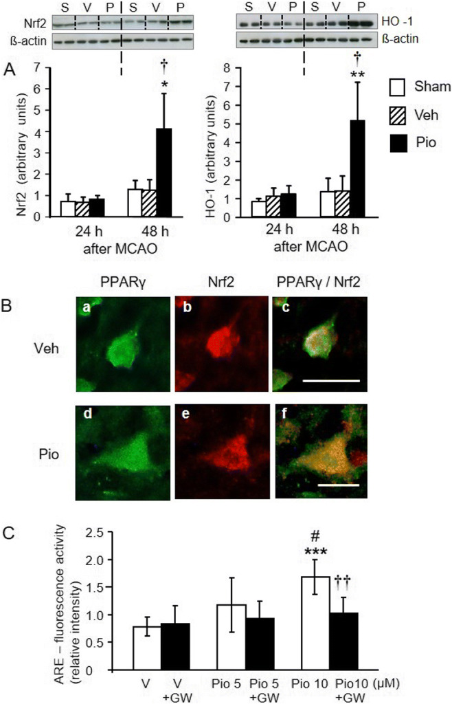
A Effects of pioglitazone (Pio) on the expression of Nrf2 (left panel) and heme oxygenase-1 (HO-1) (right panel) in the peri-infarct frontoparietal cortex 24 h and 48 h after MCAO. Pioglitazone considerably increased the expression of Nrf2 and HO-1 48 h after MCAO, when compared with the sham-operated or vehicle (Veh)-treated groups of rats. Representative blots and graphical analysis are shown for each parameter (S, sham; V, vehicle-treated; P, pioglitazone-treated rats). Results are expressed as the means ± SD. *P<0.05; **P<0.01, statistical comparisons with sham-operated rats and †P<0.05, statistical comparison with the vehicle-treated group of rats exposed to MCAO, calculated by Kruskal-Wallis test. B Immunofluorescence staining for PPARγ (a, d) (green) and Nrf2 (b, e) (red) in neurons localized at the border of the ischemic core in vehicle (Veh)-treated (upper panels) and pioglitazone (Pio)-treated (lower panels) rats. Overlapping of PPARγ and Nrf2 immunoreactivities (c, f) (yellow) shows a considerable induction of Nrf2 in pioglitazone-treated rats. Scale bar: c 50 μm, f 25 μm. C PC12 cells were transfected with reporter construct pNQO1-r antioxidant response element (ARE) plasmid and exposed to vehicle (V) or pioglitazone (Pio). Pioglitazone activated ARE and this effect was completely abolished by GW 9662 (GW). Results are expressed as the means ± SD. ***P<0.001 statistical comparison with vehicle-treated cells; #P<0.05, statistical comparison with cells exposed to 5 μM pioglitazone. ††P<0.01, statistical comparison with cells treated with 10 μM pioglitazone, calculated by one-way ANOVA followed by a post hoc Bonferroni test
