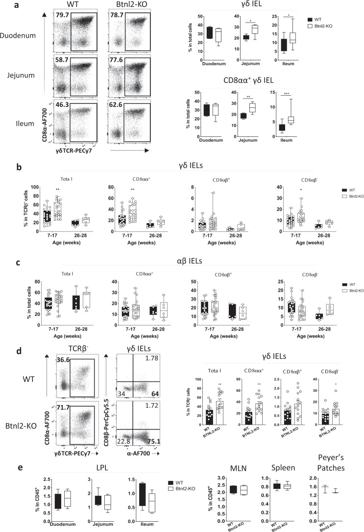
Fig. 2. Btnl2-KO mice display increased frequencies of γδ IELs in the ileum.
a Different segments of the small intestine were collected from cohoused 7–17-week-old Btnl2-KO and WT mice (n = 3–8, each) and processed for flow cytometry. Left panel-representative flow cytometry plots of γδ IELs in the duodenum, jejunum, and ileum of cohoused 7-week-old Btnl2-KO and WT littermates. Displayed plots are gated on live TCRαβ- cells. Right panel-frequencies of γδ IELs and CD8αα + γδ IELs from 7–17-week-old Btnl2-KO and WT littermates. b Frequencies of ileal γδ IELs at different ages (n = 6–23 mice/group). Error bars represent mean ± SEM. Significance is measured by 2-way ANOVA with Sidak’s multiple comparison test, *p < 0.05, **p < 0.005. c Frequencies of ileal αβ IELs at different ages (n = 6–23 mice/group). Error bars represent mean ± SEM. Significance is measured by 2-way ANOVA. d Ileum was collected from cohoused Btnl2-KO and WT littermates of different ages and intestinal intraepithelial lymphocytes (IELs) were isolated and processed for flow cytometry. Left panel-representative flow cytometry plots of γδ IELs in the ileum of 12–17-week-old Btnl2-KO and WT littermates. Right panel-frequencies of ileal γδ IELs from 12–17-week-old Btnl2-KO and WT littermates. Data are pooled from 3 independent experiments with 3–6 mice/group. Error bars represent mean ± SEM. Significance is measured using unpaired t-tests assuming similar SD, *p < 0.05, **p < 0.005, significantly different from WT mice. e Frequencies of γδ T cells in lamina propria (LP), mesenteric lymph nodes (MLN), spleen, and Peyer’s Patches of 7–17-week-old Btnl2-KO and WT littermates (n = 3–6 mice/group).

