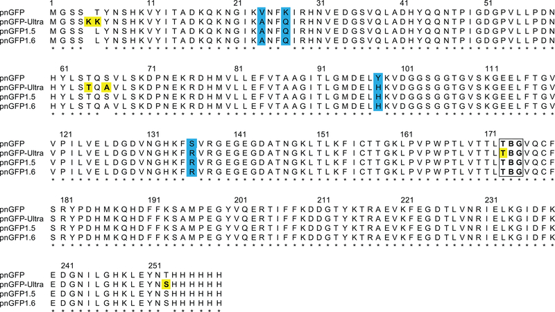
Figure 2. Sequence Alignment of pnGFP-Ultra and Related Proteins.
Mutations identified during directed evolution are highlighted in cyan. Residues subjected to rational mutagenesis are yellow-colored. The chromophore-forming residues (B denoting pBoF) are highlighted in a box. Residues are numbered according to the numbering of pnGFP1.5-Y.Cro (PDB 5F9G).

