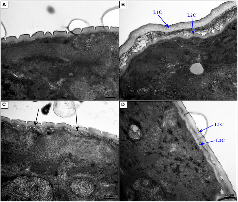
FIGURE 6.
Transmission electron microscopy of nas-33 and gpb-1 RNA interference-treated Haemonchus contortus. (A) Cuticle structure of Bt-Cry1AC RNAi-treated worm. (B) Two layers of cuticles of nas-33 RNAi-treated worm. (C) Loose connection between cuticle and epidermis in gpb-1 RNAi-treated worm. (D) Two layers of cuticles and loose connection between cuticle and epidermis of nas-33 and gpb-1 RNAi-treated worm. L1C, L1 cuticle; L2C, L2 cuticle. Black arrows point to the loose connections between cuticle and epidermis. Scale bar: 0.5 μm.

