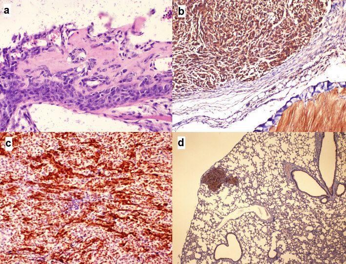
Figure 4.
The microphotographs of the morphological and immunohistochemical experiments. a. A microscopic focus showing minimal differentiation of osteosarcoma cells characterized by osteoid matrix production (hematoxylin-eosin staining; 400). b. Osteosarcoma cells displaying diffuse cytoplasmic staining with BMP2 antibody, similar to the striated muscle bundles on the right lower corner of the frame (immunohistochemistry staining; 100). c. Osteosarcoma cells displaying score 12 nuclear and cytoplasmic staining patterns with p27 (immunohistochemistry staining; 100). d. 1 (+) staining intensity similar to bronchial epithelium with caspase-3 displayed in a metastatic focus in lung tissue (Immune histochemistry staining; 40).

