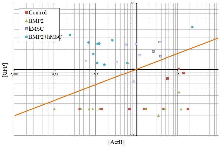
Figure 5.

GFP expression in vivo. Screening of GFP expression in the tissue after the transplantation of hMSCs and BMP2+hMSC. The GFP expression could be detected in the samples above the orange line, where only the cell transplanted groups (hMSC and BMP2+hMSC) were localized. No significant GFP expression was measured in the control group and BMP2 groups.
