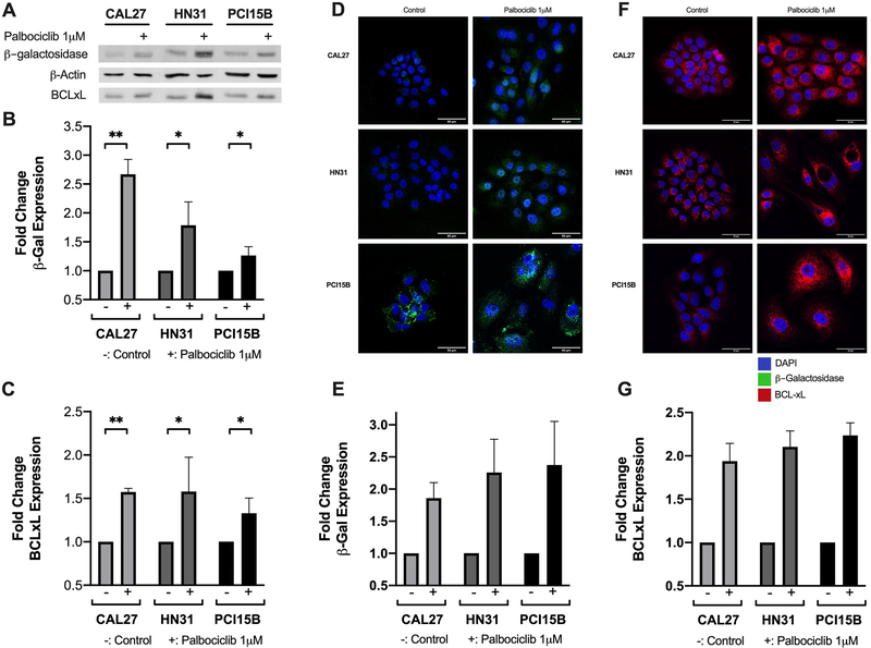
Figure 3.
β-galactosidase and BCL-xL expression following palbociclib treatment among the panel of HPV- head and neck squamous cell carcinoma (HNSCC) lines. Representative western blot showing protein expression for β-galactosidase (β-gal), BCL-xL, and β-actin, with (+) designating treatment condition with 1 μM palbociclib (A). Fold changes shown in β-gal (B) and BCL-xL (C) relative to untreated controls. HPV-lines were exposed to 1 μM palbociclib for 24 hours prior to immunofluorescent staining. Representative epifluorescence microscopy images shown for both β-gal (D) and BCL-xL (F), with DAPI in blue, β-gal in green, and BCL-xL in red. Fold changes in β-gal (E) and BCL-xL (G) relative to untreated controls. Scale bar indicates 50μm. Data in (B) and (C) displayed as mean ± standard deviation; data in (E) and (G) displayed as mean ± standard error of the mean. All data represent at least three biological replicates performed on different days. Statistical significance is indicated: * if p < 0.05, ** if p < 0.01, comparing treatment group with control group using unpaired t-test.

