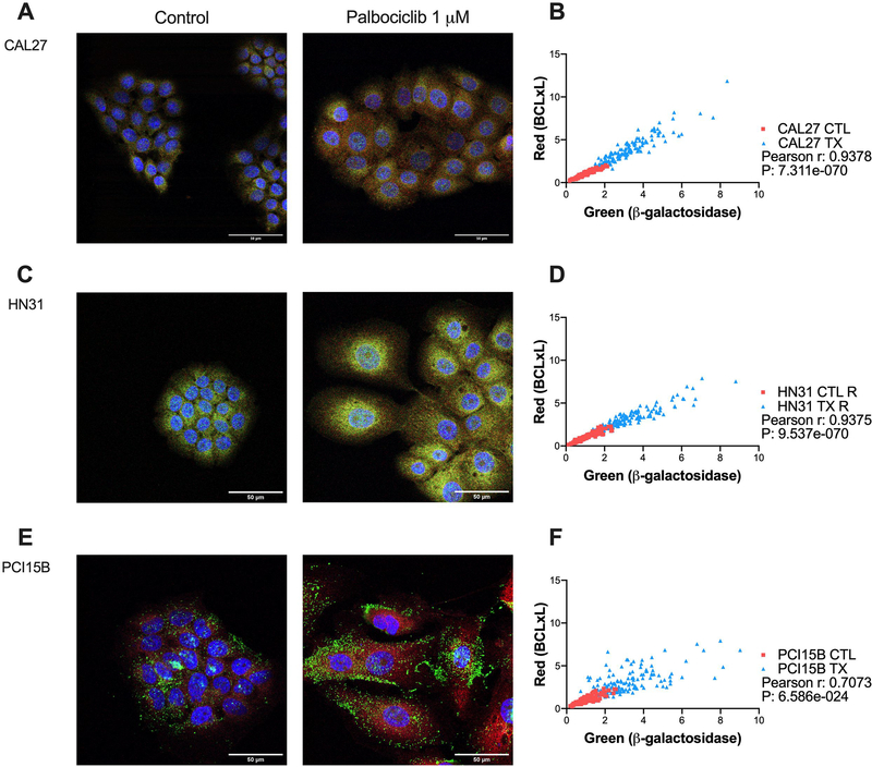
Figure 4.
Co-expression of β-galactosidase and BCL-xL following palbociclib treatment among the panel of HPV- head and neck squamous cell carcinoma (HNSCC) lines. Cells were exposed to 1 μM palbociclib for 24 hours before immunofluorescent staining. Representative confocal microscopy images for control and treatment conditions shown (A, C, E), with DAPI in blue, β-gal in green, and BCL-xL in red. Co-expression analysis for each cell line (B, D, F), with every point on the graph representing the pixel intensity per cell for one cell for β-gal and BCL-xL, and overall illustrating strong correlation for all lines. Scale bar indicates 50μm. Data displayed from three biological replicates performed on different days.

