



Abstract
Introduction:
In this study we explored walking activity in a large cohort of boys with Duchenne muscular dystrophy (DMD).
Methods:
Step activity (monitored for 7 days), functional ability, and strength were quantified in ambulatory boys (5-12.9 years of age) with DMD and unaffected boys. Ambulatory status was determined 2 years later.
Results:
Two to 5 days of activity monitoring predicted weekly step activity (adjusted R2 = 0.80-0.95). Age comparisons revealed significant declines for step activity with increasing age, and relationships were found between step activity with both function and strength (P < .01). Our regression model predicted 36.5% of the variance in step activity. Those who were still ambulatory after 2 years demonstrated baseline step activity nearly double that of those who were no longer walking 2 years later (P < .01).
Discussion:
Step activity for DMD is related to and predictive of functional declines, which may be useful for clinical trials.
Keywords: accelerometry, daily steps, Duchenne muscular dystrophy, functional ability, physical activity, walking
1 |. INTRODUCTION
Step activity and community ambulation data have recently been recognized as being both valid and sensitive real-world measures that can be of utility as outcome measures in clinical trials for Duchenne muscular dystrophy (DMD).1 In unaffected individuals, walking and step activity are important variables for assessing fitness and overall health.2–5 Accelerometry is often employed to assess step activity6–8; however, one must know the needed time to measure step activity to obtain a reasonable estimate of overall activity for a specific patient population.9,10
Although step activity in DMD has been studied,1,11,12 the minimal number of days required to best represent activity levels of boys with DMD has not been investigated. Seven consecutive days of activity monitoring has been recommended,9,10,13–16 but that amount may be viewed as a participant burden and decrease adherence/compliance in clinical studies. The number of days for step activity monitoring has varied in previous DMD studies.1,11,12,17 The aforementioned studies provide useful information regarding step activity in DMD, but it is not known whether a sufficient number of days were monitored.
Data on how age and disease progression impact activity levels in DMD also are lacking. The aforementioned studies included sample sizes ranging from 16 to 42 participants.1,11,12,17 Two of the studies used a cross-sectional design to compare activity between boys with DMD and healthy controls.11,12 The other two studies were longitudinal in nature and examined activity data across a range of age values.1,17 However, a statistical exploration of the variation in step activity data between multiple age groups for DMD is still needed.
The primary objectives in this study of a large cohort of boys with DMD were to: (a) determine the necessary time frame to obtain consistent estimates of step activity; (b) statistically assess the step activity across multiple age groups; (c) examine how step activity can be explained by functional abilities and strength; and (d) compare step activity levels for boys with DMD who remained ambulatory 2 years after accelerometry assessment vs those who became nonambulatory. Secondary objectives included the comparison of step activity in boys with DMD to unaffected controls and describing the relationships between step activity, functional abilities, and strength.
2 |. METHODS
2.1 |. Subjects
Ambulatory boys with DMD and unaffected control boys were recruited as part of a cross-sectional, multicenter, natural history study (ImagingDMD) conducted at the University of Florida, Oregon Health & Science University, and Children’s Hospital of Philadelphia. Inclusion criteria for all subjects were being 5.0 to 12.9 years old and not participating in organized sports more than twice per week. Additional inclusion criteria for the DMD participants were being able to walk 100 meters independently and ascend four steps. Boys with DMD who were participating in a clinical trial were excluded. Reports confirming the diagnosis of DMD using molecular genetic testing was obtained from each of the DMD participants. For every participant in the study a legal guardian gave written consent, and each subject provided written assent. All aspects of this research project were approved by the institutional review board at the three participating institutions.
2.2 |. Activity monitoring
Both DMD and control group participants were provided with an accelerometer (GT3X Actigraph, Actigraph Corporation, Pensacola, Florida) during one of their ImagingDMD visits and shown how to properly position and wear the device on the right side of their waists. Participants were asked to wear the device for all waking hours (except for bathing and swimming) for 7 consecutive days upon returning home. Seven days is the time frame others have recommended when investigating the duration needed for a consistent measure of step activity.9,10,13,16 The participants and their families were also provided with written instructions regarding the positioning and care of the accelerometer. To enhance compliance, reminder phone calls were made to the participants’ homes the day after step activity monitoring was to begin and again 7 days later. Once the participants had completed the step activity monitoring, the devices were returned by prepaid mail. The data were uploaded via ActiLife software (ActiGraph, Actigraph Corporation, Pensacola, FL) to be analyzed, and bilateral daily step count was calculated for step activity. The minimum amount of wear time for the accelerometer to be considered acceptable for data analyses was 10 h/day.18–23 In addition, we examined the ambulatory status of the boys with DMD who had participated in the step activity data collection at an ImagingDMD visit 2 years later. Subjects were classified as “ambulatory” if they were able to independently walk 10 meters without any assistive device or braces.
2.3 |. Functional ability
At the same ImagingDMD visit when accelerometers were dispensed, all subjects performed three timed functional tasks: 10-meter walk/run; ascend four stairs (4 stairs); and rise from the floor (Supine Up).24 Subjects performed each task three times while wearing footwear, and the fastest time was recorded for analysis. In addition, subjects completed the 6MWT with the distance walked on the 6MWT (6MWD) as the outcome measure for this test.25
2.4 |. Strength
Isometric peak torque of the knee extensor (KE) and plantar flexor (PF) muscle groups was assessed in all participants for the right leg using a dynamometer (Biodex System 4 Pro, Biodex Medical Systems, Shirley, NY), as described elsewhere.26–29 For KE testing, the hip and upper trunk were secured with straps to the dynamometer, and the hip and knee joint angles were set at 90°. The subject was instructed to push as hard as possible for 5 seconds, with 1 minute of rest between trials. PF testing used a custom-built platform where the subject was secured within a seat upon the platform by straps at the torso and the leg. The knee was placed between 20° and 30° of flexion and the ankle was placed in the neutral position (0° of plantar flexion). The subject was instructed to push on the footplate as hard as possible for 5 seconds, followed by a 1-minute rest. A minimum of five trials were performed; in the event that the last trial’s torque output exceeded all of the previous trials, the subject was asked to perform additional trials until there was a decrement or plateau in force production. The highest torque value normalized to body weight (Nm/kg) was used for analysis (for both KE and PF peak torque).
2.5 |. Statistical tests
The Shapiro-Wilk test confirmed normal distributions of the step activity and 6MWD data, and multiple linear regression models were run on the subset of DMD participants who wore the accelerometer for 7 consecutive days to examine the number of days of wear needed to predict 7 days of step activity. We followed similar methods utilized by others when investigating minimum number of days needed to obtain consistent estimates of step activity in other patient populations.10 In brief, the mean of 7 days of step activity was the dependent variable, and the independent variables were all the possible combinations of the days of the week. This resulted in 42 combinations for the variable of step activity. Independent-samples t tests were used to assess differences in step activity between boys with DMD and unaffected control subjects as well as between participants still able to walk after 2 years and those who had become nonambulatory. One-way analyses of variance with Bonferroni post hoc testing was run for comparisons between age groups and DMD subjects with different 6MWDs (0-300, 301-400, and >400 meters).30 Participants unable to complete the Supine Up were assigned the highest value for that test, so Spearman correlations were used to examine the relationship for DMD boys between step activity, functional abilities, and strength.31 Mann-Whitney U tests were performed to examine the differences between boys with DMD and controls for strength and function. Stepwise multiple linear regression models were also run to examine how functional abilities and strength explain step activity in boys with DMD. In this prediction model, step activity was the dependent variable. The independent variables were: performance in the three functional tests (10-meter walk/run, 4 stairs, and Supine Up); peak torque values for the KE and PF; and the 6MWD. Significance for all testing was set at α = 0.05.
3 |. RESULTS
Seventy ambulatory boys with DMD and 10 unaffected control boys participated in our study. Table S1 contains the genetic mutation information for the DMD participants. Table 1 demonstrates that, although there was no difference between the groups in body mass index, boys with DMD weighed less and were shorter. Unsurprisingly, the boys with DMD walked a shorter distance on the 6MWT, took longer to complete the functional tasks, and demonstrated lower leg strength relative to the controls.
TABLE 1.
Demographics of study participants
| DMD group | Control group | |
|---|---|---|
| Number of participants | 70 | 10 |
| Age, years | 8.7 (2.0) | 9.2 (1.3) |
| Weight, kg | 61.5 (19.5) | 77.2 (17.8)* |
| Height, cm | 122 (11) | 141 (7)** |
| Body mass index, kg/m2 | 18.5 (3.2) | 17.5 (2.2) |
| Participants taking steroids, % (n) | 88.5% (62) | – |
| Prednisone, % (n) | 57.1% (40) | – |
| Deflazacort, % (n) | 31.4% (22) | – |
| 6MWD (m) | 368 (64) | 599 (63)** |
| 10-meter walk/run (s) | 5.8 (1.9) | 3.0 (0.3)** |
| 4 stairs (s) | 4.2 (3.1) | 1.5 (0.2)** |
| Sup Up (s) | 5.3 (3.4) | 1.7 (0.5)** |
| KE peak torque (Nm/kg) | 0.61 (0.41) | 2.56 (0.66)** |
| PF peak torque (Nm/kg) | 1.25 (0.36) | 2.79 (0.72)** |
Note: Data expressed as mean (standard deviation).
4 Stairs, time to ascend 4 stairs; 6MWD, distance walked on 6-minute walk test; 10-meter walk/run, time to traverse 10 meters; DMD, Duchenne muscular dystrophy; KE peak torque, normalized knee extensor strength; PF peak torque, normalized plantar flexor strength; Sup Up, time-to-rise-from-floor test.
P < .05 and
P < .01 for difference between groups.
Fifty of the 70 boys with DMD and all of the unaffected controls wore the accelerometer for 7 consecutive days for at least 10 hours/day. Table S2 shows the step activity of these boys for each of the 7 days of the week. We used the data from these 50 boys to run multiple linear regression models in determining the number of days needed to predict 7 days of step activity monitoring. The results demonstrate an adjusted R2 ≥ 0.80 for any combination of 2 days (weekend days or weekdays), R2 ≥ 0.85 for any combination of 3 days, R2 ≥ 0.90 for any combination of 4 days, and R2 ≥ 0.95 for any combination of 5 days. Because of these findings, we used data from all 70 boys with DMD who had worn the accelerometer for at least 5 days for the remaining analyses.
On average, the 70 subjects with DMD took 63% of daily steps compared with unaffected control boys (mean 5147 [standard deviation 2145] vs 8138 [standard deviation 2264] steps, P < .01). To examine step activity across ages, the DMD subjects were further divided into four groups based on 2-year age ranges, as described elsewhere24: 5 to 6.9 years (n = 18); 7 to 8.9 years (n = 20); 9 to 10.9 years (n = 20); and 11 to 12.9 years (n = 12). An overall decline in daily step count was noted with increasing age, with the three older age groups being significantly different from the 5- to 6.9-year-olds (Figure 1). Significant relationships were found between step activity and functional abilities as well as strength (Figure 2). Supine Up, 10-meter walk/run, and 4 stairs were the functional tests with the greatest correlation coefficients with step activity. The multiple regression results for the prediction of step activity from functional abilities and strength of the 70 boys with DMD were statistically significant. An adjusted R2 of 0.365 (P < .001) was found, demonstrating that 36.5% of the variance in step activity is accounted for by a combination of the six strength and functional tests entered into the multiple regression model. From the independent variables, the 10-meter walk/run was the only significant predictor (standardized coefficient) of step activity.
FIGURE 1.
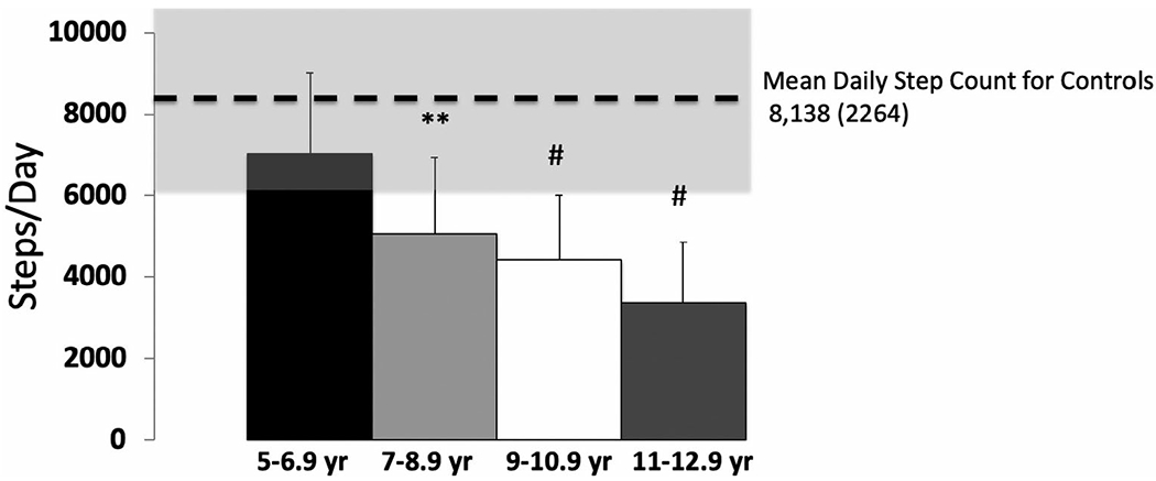
Comparison of step activity in steps per day (mean ± standard deviation) across four age groups from a large sample of boys with Duchenne muscular dystrophy (n = 70): 5 to 6.9 years (n = 18) = 7033 ± 1957 steps; 7 to 8.9 years (n = 20) = 5055 ± 1889 steps; 9 to 10.9 years (n = 20) = 4443 ± 1587 steps; and 11 to 12.9 years (n = 12) = 3385 ± 1522 steps. Dashed line represents the mean and gray area represents the standard deviation for the step activity of 10 healthy control participants. 6MWD, 6-minute walk test distance; KE, knee extensor muscle group; PF, plantar flexor muscle group
FIGURE 2.

Relationship between step activity, functional abilities, and strength in boys with DMD. A, 6MWD, distance walked for 6-minute walk test. B, Ten-meter walk/run, time to traverse 10 meters. C, Four stairs, time to ascend 4 steps. D, Supine Up, time to rise from the floor. E, KE peak torque, normalized knee extensor strength. F, PF peak torque, normalized plantar flexor muscle strength. Spearman correlations used for all variables. **Correlation significant at the P < 0.01 level
Step activity was positively correlated with 6MWD (Figure 2A), and we examined these variables further in groups of boys with DMD stratified by 6MWD. Those with a 6MWD over 300 meters were significantly different in step activity compared with those with a 6MWD less than 300 meters (Figure 3A). Thirty-six of the 70 participants with step activity data collected were seen 2 years later for an ImagingDMD visit. Of those 36, 11 were no longer ambulatory. Those who were still ambulatory after 2 years had taken, on average, 98% more daily steps during the first data collection period than those who were no longer able to walk (Figure 3B).
FIGURE 3.

Comparison of step activity (mean steps/day ± SD) across: A, Three groups of boys with DMD based upon the distance walked in the 6-minute walk test: 0 to 300 meters (n = 9) = 3170 ± 1782; 301 to 400 meters (n = 33) = 5388 ± 1764; >400 meters (n = 19) = 5806 ± 2515. B, Two groups of boys with DMD, those who lost the ability to ambulate within 2 years (n = 11) = 3112 ± 1166 steps, and those who were still walking 2 years after assessing step activity (n = 25) = 6162 ± 2290 steps. Amb, ambulant (those still walking); LOA, lost ability to ambulate
4 |. DISCUSSION
Our data for step activity demonstrate that any 2 days (weekend days or weekdays) of step activity data collection estimate the average weekly amount when wearing the accelerometer for at least 10 hours/day. This finding is based on an adjusted R2 value of 0.80, which is considered adequate for prediction purposes.10 However, for investigative purposes, when a stronger prediction is needed, we decided to provide data up to an R2 value of 0.95, requiring 5 days of step activity data collection (regardless of whether weekend days or weekdays are included). This more conservative value is what we used for our subsequent analyses combining all 70 participants. Although there has been some initial investigation into step activity in DMD,1,11,12 this information on the varying predictive strengths of different time frames of step activity assessment will be useful for future clinical trials examining how step activity is influenced by therapeutic interventions. It may also guide assessment protocols, reduce the burden on participants, and potentially improve participant adherence and compliance in future clinical studies.13
Another contribution from this work is the statistical comparison of step activity across multiple age groups. Our findings of a decrease in step count support those of Fowler et al,1 who they projected longitudinal trends from participants 4 to 16.5 years of age. Relative to the group 5 to 6.9 years old, the older age groups in our study were 72% (7-8.9 years), 63% (9-10.9 years), and 48% (11-12.9 years) as active. These percentage decreases are quite consistent with those of Fowler et al1 from a similar age range. The average amount and variability of step activity for the four age groups outlined in this study can be used for comparison purposes in future clinical trials that focus on increasing step count or prolonging ambulatory ability in boys with DMD.
Furthermore, this study’s large sample of boys with DMD allowed the use of regression to predict step activity from functional abilities and strength. Based on the standardized coefficient, the 10-meter walk/run was the most important independent predictor for step activity. Fowler and colleagues1 also described the importance of the 10-meter walk/run test as it relates to step activity measures. Our group recently described the value of the 10-meter walk/run test as a highly sensitive measure with a strong predictive value for assessing ambulatory function in boys with DMD.24 Having knowledge about how patients perform on the 10-meter walk/run test may be useful to clinicians and scientists when estimating step activity for ambulatory boys with DMD. Our results suggest other factors exist beyond functional abilities and strength that are important to predict step activity in these boys, such as psychosocial aspects, parental perception of benefit or harm from step activity, fear of falling, etc.32–34 Further work should strive to describe these factors and how they also contribute to step activity in this patient population.
The 6MWT has also been shown to have a significant relationship for r2 with step activity.12 We divided our participants into three groups based on 6MWD30 to examine step activity. There was an apparent difference most noted at a 6MWD of 300 meters. When we examined who lost the ability to ambulate 2 years after step activity data collection, those 11 participants had a mean step activity of only ~50% relative to the group that was still ambulatory (Figure 3). It is of interest how similar the step activity is between those with a 6MWD of 0 to 300 meters and those who lost ambulatory ability within 2 years (a difference of <2%). Others investigators have proposed important cutoff values for the 6MWD of 325 and 350 meters for estimating rate of functional decline and/or loss of ambulation in DMD.35 Similar 6MWD values have been used in clinical trials for inclusion and stratification criteria in clinical trials.36 Our data support these cutoff values for the 6MWD and also provide a potential cutoff value for step activity estimating loss of ambulation within 2 years. This additional information may also prove useful for clinical trial recruitment, screening, and implementation.
Our control group participants were less active than the healthy children in an earlier study,37 and our exclusion of unaffected boys who participated in organized sports more than twice/week may have excluded some of the more active population. The boys with DMD in this study only took 63.2% of the daily steps relative to our controls. These findings support those of others who used smaller sample sizes (n = 16 boys with DMD) and reported daily steps in boys with DMD to be approximately 70% of that taken by unaffected boys.11 We found the strongest relationship between step activity and timed functional tests (particularly the 10-meter walk/run) and moderate correlations with 6MWD, KE peak torque, and PF peak torque. Our findings are similar to those of another study reporting a relationship between step activity and 6MWD.12 However, our results differ from those Fowler et al1 and McDonald et al,11 who did not find a significant relationship between steps per day with 10-meter walk/run speed1 and KE strength (normalized to body mass),11 respectively. These differing findings are most likely attributed to smaller samples in the other studies and thus did not have the power of the current study with its larger cohort to detect statistical significance.
This study does have some limitations. First, the accelerometer used was a waist-worn device. Earlier studies of step activity for DMD1,11,12 utilized ankle-worn accelerometers, which may be more precise in assessing step activity.3 The step activity results from our study, however, are comparable with what others have reported using ankle-worn devices. In addition, the accelerometer has been widely used to assess step activity in various patient populations, with valid and reliable results.38–44 A second limitation is that these data came from participants in a large natural history study. Therefore, there was no control of clinical care or medications, and this medical management depended on the recommendations and prescriptions of the physician(s) for each participant. However, due to this varied clinical care and medication regimens as well as the fact that none of the 70 participants were participating in a clinical trial during this study, our findings should be more generalizable to this patient population as a whole.
In conclusion, our study has provided valuable information on step activity in DMD. The results show varying degrees of strength from different time periods of step activity monitoring to estimate community walking. Our findings also demonstrate statistical differences and variability for step activity across various age groups that can be used as reference data for future studies. The utility of the 10-meter walk/run as a test of ambulatory function for boys with DMD is reinforced, and a potential cutoff value for step activity in predicting loss of ambulation is provided. Collectively, these results should assist clinicians, scientists, and therapeutic companies in the design and implementation of future clinical trials focused on walking ability in boys with DMD.
Supplementary Material
ACKNOWLEDGMENTS
The ImagingDMD study was financially supported by funding from the National Institute of Arthritis and Musculoskeletal and Skin Diseases and the National Institute of Neurological Disorders and Stroke of the National Institutes of Health (R01 AR056973). The authors are grateful to the boys who participated and the support of their families. We also thank Venessa Bibeau, Alex Millette, and Paul Park for their assistance with data processing and analyses.
Funding information
National Institute of Arthritis and Musculoskeletal and Skin Diseases and the National Institute of Neurological Disorders and Stroke of the National Institutes of Health, Grant/Award Number: R01 AR056973; Rehabilitation Research Career Development Program (NIH), Grant/Award Number: K12-HD055929
Abbreviations:
- DMD
Duchenne muscular dystrophy
- KE
knee extensor
- LOA
loss of ambulatory ability after 2 years
- PF
plantar flexor
- Supine Up
rise-from-the-floor timed test
- 4 stairs
ascend-four-stairs timed test
- 6MWD
6-minute walk test distance
- 6MWT
6-minute walk test
Footnotes
CONFLICT OF INTEREST
The authors declare no potential conflicts of interest.
ETHICAL PUBLICATION STATEMENT
We confirm that we have read the Journal’s position on issues involved in ethical publication and affirm that this report is consistent with those guidelines.
SUPPORTING INFORMATION
Additional supporting information may be found online in the Supporting Information section at the end of this article.
REFERENCES
- 1.Fowler EG, Staudt LA, Heberer KR, et al. Longitudinal community walking activity in Duchenne muscular dystrophy. Muscle Nerve. 2018;57:401–406. [DOI] [PubMed] [Google Scholar]
- 2.Tudor-Locke C, Bassett DR Jr. How may steps/day are enough? Preliminary pedometer indices for public health. Sports Med. 2004;34:1–8. [DOI] [PubMed] [Google Scholar]
- 3.McDonald CM, Widman L, Abresch RT, Walsh SA, Walsh DD. Utility of a step activity monitor for the measurement of daily ambulatory activity in children. Arch Phys Med Rehabil. 2005;86:793–801. [DOI] [PubMed] [Google Scholar]
- 4.Soroush A, Der Ananian C, Ainsworth BE, et al. Effects of a 6-month walking study on blood pressure and cardiorespiratory fitness in U.S. and Swedish adults: ASUKI step study. Asian J Sports Med. 2013;4:114–124. [DOI] [PMC free article] [PubMed] [Google Scholar]
- 5.Tam KM, Cheung SY. Validation of electronic activity monitor devices during treadmill walking. Telemed J Environ Health. 2018;24:782–789. [DOI] [PubMed] [Google Scholar]
- 6.Dyrstad SM, Hansen BH, Holme IM, Anderssen SA. Comparison of self-reported versus accelerometer-measured physical activity. Med Sci Sports Exerc. 2014;46:99–106. [DOI] [PubMed] [Google Scholar]
- 7.Prince SA, Adamo KB, Hamel ME, Hardt J, Gorber SC, Tremblay M. A comparison of direct versus self-report measures for assessing physical activity in adults: a systematic review. Int J Behav Nutr Phys Act. 2008;5:56. [DOI] [PMC free article] [PubMed] [Google Scholar]
- 8.Yano S, Koohsari MJ, Shibata A, et al. Comparison of older and newer generation active style pro accelerometers in physical activity and sedentary behavior surveillance under a free-living environment. Int J Environ Res Public Health. 2019;16:E1597. [DOI] [PMC free article] [PubMed] [Google Scholar]
- 9.Tudor-Locke C, Bassett DR, Shipe MF, McClain JJ. Pedometry methods for assessing free-living adults. J Phys Act Health. 2011;8:445–453. [DOI] [PubMed] [Google Scholar]
- 10.Almeida GJ, Wasko MC, Jeong K, Moore CG, Piva SR. Physical activity measured by the SenseWear armband in women with rheumatoid arthritis. Phys Ther. 2011;91:1367–1376. [DOI] [PMC free article] [PubMed] [Google Scholar]
- 11.McDonald CM, Widman LM, Walsh DD, Walsh SA, Abresch RT. Use of step activity monitoring for continuous physical activity assessment in boys with Duchenne muscular dystrophy. Arch Phys Med Rehabil. 2005;86:802–808. [DOI] [PubMed] [Google Scholar]
- 12.Davidson ZE, Ryan MM, Kornberg AJ, Walker KZ, Truby H. Strong correlation between the 6-minute walk test and accelerometry functional outcomes in boys with Duchenne muscular dystrophy. J Child Neurol. 2015;30:357–363. [DOI] [PubMed] [Google Scholar]
- 13.Dillon CB, Fitzgerald AP, Kearney PM, et al. Number of days required to estimate habitual activity using wrist-worn GENEActiv accelerometer: a cross-sectional study. PLoS One. 2016;11:e0109913. [DOI] [PMC free article] [PubMed] [Google Scholar]
- 14.Wentz EE, Bjornson KF, Kerfeld CI, Cicirello N, Fiss AL. Walking performance, physical activity, and validity of the early activity scale for endurance in young children with cerebral palsy. Phys Occup Ther Pediatr. 2020;40(5):557–570. [DOI] [PubMed] [Google Scholar]
- 15.Kang M, Bjornson K, Barreira TV, Ragan BG, Song K. The minimum number of days to establish reliable physical activity estimates in children aged 2-15 years. Physiol Meas. 2014;35:2229–2237. [DOI] [PubMed] [Google Scholar]
- 16.Telford RD, Cunningham RB, Telford RM. Day-dependent step-count patterns and their persistence over 3 years in 8-10-year-old children: the LOOK project. Ann Hum Biol. 2009;36:669–679. [DOI] [PubMed] [Google Scholar]
- 17.Kimura S, Ozasa S, Nomura K, Yoshioka K, Endo F. Estimation of muscle strength from actigraph data in Duchenne muscular dystrophy. Pediatr Int. 2014;56:748–752. [DOI] [PubMed] [Google Scholar]
- 18.Troiano RP, Berrigan D, Dodd KW, Masse LC, Tilert T, McDowell M. Physical activity in the United States measured by accelerometer. Med Sci Sports Exerc. 2008;40:181–188. [DOI] [PubMed] [Google Scholar]
- 19.Rich C, Geraci M, Griffiths L, Sera F, Dezateux C, Cortina-Borja M. Quality control methods in accelerometer data processing: defining minimum wear time. PLoS One. 2013;8:e67206. [DOI] [PMC free article] [PubMed] [Google Scholar]
- 20.Hawkins MS, Storti KL, Richardson CR, et al. Objectively measured physical activity of USA adults by sex, age, and racial/ethnic groups: a cross-sectional study. Int J Behav Nutr Phys Act. 2009;6:31. [DOI] [PMC free article] [PubMed] [Google Scholar]
- 21.Chen S, Kim Y, Gao Z. The contributing role of physical education in youth’s daily physical activity and sedentary behavior. BMC Public Health. 2014;14:110. [DOI] [PMC free article] [PubMed] [Google Scholar]
- 22.Wennman H, Pietilä A, Rissanen H, et al. Gender, age and socioeconomic variation in 24-hour physical activity by wrist-worn accelerometers: the FinHealth 2017 survey. Sci Rep. 2019;9:6534. [DOI] [PMC free article] [PubMed] [Google Scholar]
- 23.Kuenze C, Cadmus-Bertram L, Pfieffer K, et al. Relationship between physical activity and clinical outcomes after ACL reconstruction. J Sport Rehabil. 2019;28:180–187. [DOI] [PubMed] [Google Scholar]
- 24.Arora H, Willcocks RJ, Lott DJ, et al. Longitudinal timed functional tests in Duchenne muscular dystrophy: imaging DMD cohort natural history. Muscle Nerve. 2018;58:631–638. [DOI] [PMC free article] [PubMed] [Google Scholar]
- 25.McDonald CM, Henricson EK, Han JJ, et al. The 6-minute walk test as a new outcome measure in Duchenne muscular dystrophy. Muscle Nerve. 2010;41:500–510. [DOI] [PubMed] [Google Scholar]
- 26.Akima H, Lott D, Senesac C, et al. Relationships of thigh muscle contractile and non-contractile tissue with function, strength, and age in boys with Duchenne muscular dystrophy. Neuromuscul Disord. 2012;22:16–25. [DOI] [PMC free article] [PubMed] [Google Scholar]
- 27.Batra A, Harrington A, Lott DJ, et al. Two-year longitudinal changes in lower limb strength and its relation to loss in function in a large cohort of patients with Duchenne muscular dystrophy. Am J Phys Med Rehabil. 2018;97:734–740. [DOI] [PMC free article] [PubMed] [Google Scholar]
- 28.Arpan I, Willcocks RJ, Forbes SC, et al. Examination of effects of corticosteroids on skeletal muscles of boys with DMD using MRI and MRS. Neurology. 2014;83:974–980. [DOI] [PMC free article] [PubMed] [Google Scholar]
- 29.Mathur S, Lott DJ, Senesac C, et al. Age-related differences in lower-limb muscle cross-sectional area and torque production in boys with Duchenne muscular dystrophy. Arch Phys Med Rehabil. 2010;91:1051–1058. [DOI] [PMC free article] [PubMed] [Google Scholar]
- 30.Pane M, Mazzone ES, Sivo S, et al. The 6 minute walk test and performance of upper limb in ambulant Duchenne muscular dystrophy boys. PLoS Curr. 2014;6. https://pubmed.ncbi.nlm.nih.gov/25642376/ [DOI] [PMC free article] [PubMed] [Google Scholar]
- 31.Arpan I, Forbes SC, Lott DJ, et al. T2 mapping provides multiple approaches for the characterization of muscle involvement in neuromuscular diseases: a cross-sectional study of lower leg muscles in 5-15-year-old boys with Duchenne muscular dystrophy. NMR Biomed. 2013;26:320–328. [DOI] [PMC free article] [PubMed] [Google Scholar]
- 32.Colvin MK, Poysky J, Kinnett K, et al. Psychosocial management of the patient with Duchenne muscular dystrophy. Pediatrics. 2018;142 (suppl 2):S99–S109. [DOI] [PubMed] [Google Scholar]
- 33.Hong Y, Wang Y, Zhang Y, et al. Risk factors for falls among boys under 18 years with muscular dystrophy. J Pediatr Rehabil Med. 2019;12:3–10. [DOI] [PubMed] [Google Scholar]
- 34.Bothwell JE, Dooley JM, Gordon KE, MacAuley A, Camfield PR, MacSween J. Duchenne muscular dystrophy—parental perceptions. Clin Pediatr (Phila). 2002;41:105–109. [DOI] [PubMed] [Google Scholar]
- 35.McDonald CM, Henricson EK, Abresch RT, et al. The 6-minute walk test and other endpoints in Duchenne muscular dystrophy: longitudinal natural history observations over 48 weeks from a multicenter study. Muscle Nerve. 2013;48:343–356. [DOI] [PMC free article] [PubMed] [Google Scholar]
- 36.Bushby K, Finkel R, Wong B, et al. Ataluren treatment of patients with nonsense mutation dystrophinopathy. Muscle Nerve. 2014;50:477–487. [DOI] [PMC free article] [PubMed] [Google Scholar]
- 37.Colley RC, Garriguet D, Janssen I, Craig CL, Clarke J, Tremblay MS. Physical activity of Canadian children and youth: accelerometer results from the 2007 to 2009 Canadian Health Measures Survey. Health Rep. 2011;22:15–23. [PubMed] [Google Scholar]
- 38.Anderson JL, Yoward LS, Green AJ. A study investigating the validity of an accelerometer in quantification of step count in adult hospital inpatients recovering from critical illness. Clin Rehabil. 2019;33:936–942. [DOI] [PubMed] [Google Scholar]
- 39.Do VV, Jancey J, Pham NM, Nguyen CT, Hoang MV, Lee AH. Objectively measured physical activity of Vietnamese adults with type 2 diabetes: opportunities to intervene. J Prev Med Public Health. 2019;52:101–108. [DOI] [PMC free article] [PubMed] [Google Scholar]
- 40.Joschtel B, Gomersall SR, Tweedy S, Petsky H, Chang AB, Trost SG. Objectively measured physical activity and sedentary behavior in children with bronchiectasis: a cross-sectional study. BMC Pulm Med. 2019;19:7. [DOI] [PMC free article] [PubMed] [Google Scholar]
- 41.Ahmadi M, O’Neil M, Fragala-Pinkham M, Lennon N, Trost S. Machine learning algorithms for activity recognition in ambulant children and adolescents with cerebral palsy. J Neuroeng Rehabil. 2018;15:105. [DOI] [PMC free article] [PubMed] [Google Scholar]
- 42.Baque E, Barber L, Sakzewski L, Boyd RN. Reproducibility in measuring physical activity in children and adolescents with an acquired brain injury. Brain Inj. 2016;30:1692–1698. [DOI] [PubMed] [Google Scholar]
- 43.Mitchell LE, Ziviani J, Boyd RN. Variability in measuring physical activity in children with cerebral palsy. Med Sci Sports Exerc. 2015;47:194–200. [DOI] [PubMed] [Google Scholar]
- 44.O’Neil ME, Fragala-Pinkham MA, Forman JL, Trost SG. Measuring reliability and validity of the ActiGraph GT3X accelerometer for children with cerebral palsy: a feasibility study. J Pediatr Rehabil Med. 2014;7:233–240. [DOI] [PubMed] [Google Scholar]
Associated Data
This section collects any data citations, data availability statements, or supplementary materials included in this article.