
Figure 2.
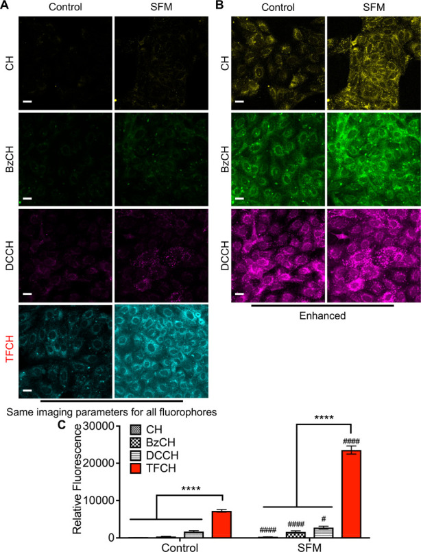
TFCH is better suited for sensing mild forms of oxidative injury in live cells. MDCK cells grown in standard media (control) or serum-free media (SFM) for 1.5 h were allowed to react with 20 μM fluorophore for 30 min, rinsed, fixed, and processed as described in the Methods. All the samples were imaged using the same imaging parameters (A). Images of the cells treated with CH, BzCH, and DCCH are enhanced for visual clarity (B). A pseudocolor was assigned to each fluorophore. Scale bar, 20 μm. Bar graphs showing quantification of cellular carbonyls detected by each fluorophore in control and serum-starved cells (C). Three independent experiments were performed, and fluorescence associated with >100 cells was quantified. An unpaired t test with Welch’s correction was performed to either compare the fluorescence signal generated by TFCH and the other fluorophores (****P < 0.0001) or to compare the fluorescence signal of each fluorophore in control and SFM treated cells (####P < 0.0001, #P < 0.05). Error bars represent SEM.